What is the Effect of Excessive Ventilation – ACLS

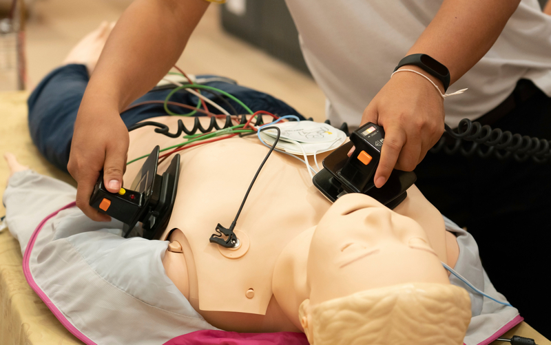
Advanced Cardiovascular Life Support (ACLS) is a set of clinical guidelines used by healthcare providers to manage cardiac arrest and other life‐threatening cardiovascular emergencies. Building on the foundation of Basic Life Support (BLS), ACLS adds critical interventions like advanced airway management, respiratory support, pharmacology, and rhythm recognition to increase a patient’s chances of survival and recovery.
But behind every successful resuscitation is a provider who understands why every second and every breath matters. When done right, ventilation ensures that oxygen is delivered to the lungs and carbon dioxide is removed, helping to preserve cellular function and minimize damage to vital organs like the heart and brain. However, when it’s done wrong, excessive ventilation during resuscitation can quickly become the difference between life and death.
Thank you for taking the time to complete this form.
What Counts as Excessive Ventilation in ACLS?
Excessive ventilation occurs when rescue breaths are delivered with too much volume, too quickly, or too frequently, surpassing the recommended ventilation rates during cardiopulmonary resuscitation. According to current ACLS guidelines, providers should aim to deliver one breath every 6 seconds (10 breaths per minute) once an advanced airway is in place. Without an airway, the recommended ratio remains 30 compressions to 2 breaths. Anything beyond these targets risks disrupting the delicate balance of circulation, respiratory function, and oxygenation.
Common Signs of Gastric Inflation and Over-Ventilation
Recognizing excessive ventilation in real time can be difficult, especially in the high-stress environment of a code. However, key indicators include:
- Visible over‐inflation of the chest, or a chest rise that is too rapid or forceful
- Gastric distension, signaling that air may be entering the stomach instead of the lungs
- Ventilations delivered faster than every 6 seconds or without full exhalation between breaths
Common Causes of Respiratory Over-Ventilation
- Anxiety and adrenaline: In high-pressure situations, it’s natural to default to faster, more forceful breaths.
- Incorrect bag-valve-mask (BVM) technique: Poor hand positioning, excessive squeezing, or lack of visual feedback can easily lead to over-delivery of air.
- Over-reliance on BVM devices: Without capnography or feedback tools, BVM ventilation can be imprecise and hard to regulate consistently.
Difference Between Adequate, Minimal, and Excessive Ventilation
 means delivering just enough air to cause visible chest rise at the correct rate—approximately one breath every 6 seconds (10 breaths per minute) with an advanced airway. The goal is to support oxygenation and carbon dioxide removal without interfering with circulation.
Minimal ventilation refers to under-ventilation—either too little air per breath or too few breaths per minute. This can lead to hypoxia and carbon dioxide buildup, both of which compromise organ perfusion and neurologic recovery.
Excessive ventilation, by contrast, occurs when breaths are delivered too forcefully, too frequently, or with too much volume. This raises intrathoracic pressure, impairs venous return, lowers cardiac output, and increases the risk of complications like gastric inflation and aspiration.
Effects of Excessive Ventilation on the Patient
When ventilation exceeds recommended rate, volume, or pressure limits. The physiological consequences can be severe and far-reaching. Even well-intentioned efforts can compromise the effectiveness of resuscitation and increase the risk of complications.
Below are the most critical effects of excessive ventilation during ACLS:
Decreased Cardiac Output
Too much air pressure in the chest makes it harder for blood to return to the heart. This reduces how much blood the heart can pump, which means less oxygen reaches the brain and other vital organs.
Altered Blood Gas Levels
Breathing too quickly blows off too much carbon dioxide, which can throw off the body’s chemical balance. This can shrink the blood vessels in the brain, reducing the oxygen it gets and worsening outcomes.
Interruptions During Chest Compressions
Focusing too much on giving breaths can lead to unnecessary pauses in CPR. These interruptions lower the chances of getting the heart started again.
Compromised Lung, Heart, and Gas Dynamics
Pushing in too much air can overfill the lungs and crowd the heart. This makes it harder for oxygen to move through the body and can make CPR less effective.
Gastric Inflation and Aspiration
Too much or too forceful breathing can send air into the stomach instead of the lungs. This raises the risk of vomiting and choking, which can make the situation worse.
Barotrauma and Alveolar Overdistention
Using too much air or squeezing the bag too hard can damage the lungs. This may cause air leaks or make it harder for the lungs to work properly after CPR.
Reduced Coronary and Cerebral Perfusion
All these issues together can reduce blood flow to the two organs that need it most during cardiac arrest: the heart and the brain.
Why Maintaining Optimal Air, Gas Exchange, and Oxygen Levels is Vital for Patient Survival
During cardiac arrest, the body’s ability to exchange gases is already compromised. That makes it even more critical to deliver oxygen efficiently and remove carbon dioxide without disrupting circulation. Proper ventilation supports this delicate respiratory balance by ensuring that oxygen reaches vital organs and waste gases are eliminated, all while allowing the heart to refill and pump effectively.
When oxygen levels drop too low, tissue hypoxia sets in, threatening the brain and heart. But delivering too much air too quickly can be just as dangerous, impairing venous return, reducing cardiac output, and causing harmful shifts in blood gas levels.
Preventing the Risks of Respiratory Complications
Preventing excessive ventilation starts with vigilance, teamwork, and adherence to ACLS best practices. Here are key strategies to reduce the risk of over-ventilating during a resuscitation event:
Capnography and Airway Monitoring
Use end-tidal CO₂ (ETCO₂) monitoring to ensure ventilation is supporting effective gas exchange.
Visual Cues and Breath Volume
Watch for visible chest rise and avoid delivering excessive volume with each breath. One breath over 1 second, just enough to make the chest rise, is sufficient. Over-inflation increases the risk of barotrauma and gastric inflation.
Proper Mask Seal and Air Delivery
Ensure a tight seal with the bag-valve-mask to direct air into the lungs, not the stomach. Poor technique can lead to air leaks or air entering the GI tract, both of which compromise effective ventilation.
Clear Communication During a Code
Assign one person to manage ventilation and ensure everyone is aware of the ventilation rate. Verbal cues like “one breath every six seconds” help maintain consistency and prevent accidental over-ventilation.
Assign the Task to the Most Trained Rescuer
Ventilation should be handled by the most experienced team member, especially in high-pressure code situations. Skilled airway management is critical to avoiding preventable complications.
Emphasis on ACLS Training
Ongoing ACLS training and simulation practice reinforce correct technique and build muscle memory.
Systematic Patient Assessment
Resuscitation is followed by a critical phase of ongoing patient assessment and stabilization. ACLS-trained providers must conduct both primary and secondary evaluations to identify immediate threats and manage post-arrest care effectively.
The primary assessment focuses on the ABCs (airway, breathing, and circulation) along with neurologic status and exposure. It ensures that the patient has a protected airway, adequate ventilation and perfusion, and no immediate life-threatening injuries. During resuscitation, this step also includes establishing vascular access and identifying shockable rhythms.
Once ROSC is achieved, a secondary assessment begins. This involves a more detailed evaluation of the patient’s history, possible causes of arrest, and targeted interventions to improve long-term outcomes.
Post-Defibrillation and Respiratory Arrest Management in ACLS
- Minimize CPR interruptions to under 10 seconds for rhythm checks or shock delivery.
- Clear the patient and remove any oxygen sources from the chest before pressing “Shock.”
- Follow AED prompts and resume compressions immediately after rhythm analysis.
After Defibrillation
- Restart CPR right away, regardless of whether a shock was delivered.
- Monitor ETCO₂—a value under 10 mmHg may indicate poor perfusion; improve compressions or consider vasopressors.
- Reassess rhythm every 2 minutes.
- Consider termination if no ROSC after 20 minutes of high-quality CPR in an intubated patient.
Certification & Training
Excessive ventilation is one of the most common mistakes made during resuscitation and one of the most avoidable. The key to prevention lies in training, practice, and up-to-date certification.
AMC’s 100% online ACLS certification and recertification courses are designed for healthcare professionals who want clinically rigorous training without the hassle of in-person classes. Whether you’re a nurse, paramedic, physician assistant, or other clinical provider, AMC’s ACLS training gives you the tools you need to master high-stakes decision-making—like proper respiratory ventilation techniques—under pressure.
Precision Saves Lives
Excessive ventilation during ACLS can undo even the most effective chest compressions and defibrillation efforts. The dangers are real and preventable with proper ventilation technique. These lifesaving skills require mastering breath rate, volume, and timing. With precision, discipline, and clinical excellence, every breath becomes a step toward your patient’s recovery.
If you’re ready to strengthen your skills and stay current with the latest ACLS protocols, AMC’s 100% online certification and recertification courses offer the flexibility, speed, and clinical accuracy you need. Get started on your ACLS training today.