Blogs – orig

Essential Insights: Normal Respiratory Rates for Kids & Adults

Essential Insights: Normal Respiratory Rates for Kids & Adults

Photo of Greta

by Greta Kviklyte

Life Saver, AMC
Co-authored by Kim Murray, RN, M.S.

posted on Dec 29, 2021, at 5:52 am

Share

In emergency situations, being able to recognize quickly whether a person is suffering from respiratory distress is critical to ensure that life-saving measures are implemented as soon as possible.

Children and adults suffering from respiratory distress will typically present with similar signs and symptoms, but there are significant differences between normal respiration in a child vs an adult. Understanding these differences will assist the professional rescuer (or bystander) in providing effective emergency care.

This article discusses important things to know about the normal respiratory rate in children, the common causes, signs, and symptoms of pediatric respiratory distress, complications associated with respiratory distress, and how to manage or treat breathing problems in a child.

But first, a review of important definitions, as well as a very brief summary of the respiratory system itself, should add some helpful context to this discussion.

Helpful Definitions

  • Respiratory failure: a condition in which blood does not have enough oxygen and/or too much carbon dioxide; can be chronic or acute
  • Respiratory distress: an emergency medical condition in which a person is struggling to breathe
  • Ventilation: the mechanical act of breathing in which air flows into the lungs (inhalation) and out of the lungs (exhalation)
  • Respiration: the physiological effect of breathing in which gas (e.g., oxygen and carbon dioxide) is exchanged between the environment and cells in the body
  • Hypoxemia: not enough oxygen in the blood
  • Hypercapnia: too much carbon dioxide in the blood

The Respiratory System 101: Parts and Process

ecg and patient monitorThe main goals of the respiratory system are to bring oxygen into the body and expel carbon dioxide out of the body. Some of the main structures involved in the respiratory system include, but are not limited to, the following:

  • Mouth and nose
  • Sinuses: hollow spaces between bones in the head that help regulate humidity and temperature of inhaled air
  • Pharynx (throat): the tube-like passageway through which air travels from the mouth and nose to the trachea
  • Trachea (windpipe): the tube-like passageway that connects the throat and lungs
  • Bronchial tubes: major tube-like passageways that are found at the bottom of the trachea and connect to each lung
  • Lungs: the two organs, divided into lobes or sections (three lobes on the right lung and two lobes on the second lung), that fill with air and allow gases to exchange between air and blood
    • Alveoli: tiny air sacs in the lungs
    • Bronchioles: smaller tubes through which air flows from the bronchial tubes to the alveoli
  • Diaphragm: a tent-like muscle beneath the lungs that helps airflow in and out of the lungs

What Happens When You Breathe?

The following is the basic process of what happens during inhalation and exhalation:

  • As a person breathes in, the diaphragm moves down and the ribs move up and out. This helps the chest area become bigger, so the lungs have room to expand. This also decreases the pressure inside the alveoli so that air flows into the lungs (air, like other gases, moves from areas of higher pressure to lower pressure).
  • Oxygen in the air moves from the alveoli into the blood flowing inside tiny blood vessels called capillaries, which are located inside the alveolar walls.
  • Once inside the blood, oxygen attaches to proteins on red blood cells called hemoglobin. The heart pumps this oxygen-rich blood around the body in blood vessels called arteries.
  • As cells and tissues in the body receive and use oxygen from the blood, they send off carbon dioxide into the blood.
  • This carbon dioxide-rich blood flows back to the heart in blood vessels called veins, and then the heart pumps this carbon dioxide-rich blood back to the lungs.
  • Carbon dioxide moves from the capillaries back into the alveoli. As a person breaths out, the diaphragm moves up and the ribs move down. This helps the chest area become smaller and increases the pressure inside the lungs, allowing air to get pushed out.

What Is a Normal Respiratory Rate for Adults?

The normal respiratory rate for adults is between 12 and 18 breaths per minute at rest. If respiration rates are under 12 breaths per minute, a person may not get enough oxygen to meet their body’s needs. Respiratory rates over 25 can indicate an underlying health condition or trouble breathing.

What is a Normal Respiratory Rate for a Newborn?

Newborns have a much faster normal respiratory rate than adults. Typically, babies will breathe at a rate of 40 to 60 times per minute at a normal breathing rate. As the baby falls asleep, their breathing rate may fall to between 30 and 40 times per minute. Breathing faster than adults is not a sign of a health complication for newborns unless it is beyond 60 times per minute. Children who are born early or with other health complications may have a breathing rate that is much outside of this range, and that could indicate underdeveloped lungs.

The Normal Respiratory Rate in Children

doctor-checking-childs-respiratory-ratehe respiratory rate refers to the number of times a person breathes in one minute while at rest (i.e., not performing exercise or physical activity). The normal respiratory rate in a child tends to be quicker than an adult’s.

Understanding the normal breathing rate for children can help a professional or bystander rescuer more quickly recognize whether a child is suffering from breathing difficulties.

Typical respiratory rates for children, based on age range, are as follows:

  • Infant (0-12 months old): 30 to 60 breaths per minute
  • Toddler (1-3 years old): 24-40 breaths per minute
  • Preschooler (4-5 years old): 22-34 breaths per minute
  • School-aged child (6-12 years old): 18-30 breaths per minute

By the time a child reaches adolescence (13 to 18 years old), their normal respiratory rate will be the same as an adult’s—about 12 to 16 breaths per minute.

Interestingly, infants and young children also tend to have faster heart rates (pulses) than adults on average. The normal resting heart rate of a newborn (0 to 3 months old) ranges from 80 to 205 beats per minute, and a child aged 4 months to 2 years has a normal resting heart rate of 75 to 190 beats per minute. Between ages 2 and 10, a child’s heart rate will slow to about 70 to 140 beats per minute.

By the time she is 10 years old, a child’s resting heart rate will be similar to an adult’s (60 to 100 beats per minute).

Why Do Children Breathe Faster Than Adults?

Generally speaking, children tend to breathe faster than adults do because they have smaller lungs (lower lung volume) than adults, and therefore have less physical space to exchange gases.

Younger children also tend to have a much faster metabolism than adults, meaning they break down nutrients at a faster rate. As a result, children typically require more oxygen (and faster disposal of carbon dioxide) while at rest compared to adults, even after adjusting for body mass and unit of time.

What Is a Low Respiratory Rate?

A low respiratory rate is typically under 12 breaths per minute for adults or under 20 breaths per minute for children. Newborns should not be below this figure, though some slightly older children may be.

A low breathing rate indicates that the body may not be getting enough oxygen to meet its needs. Too low a respiratory rate is called bradypnea, and it requires immediate medical attention.

What Is a High Respiratory Rate?

A high respiratory rate occurs when a person breathes very quickly, often not getting deep breaths. This condition, called tachypnea, can mean not enough air is getting into the lungs to meet the body’s needs. In adults, breathing rates of over 25 breaths per minute can be problematic, and in children, breathing rates over 40 breaths per minute in most children or 60 breaths per minute in newborns can indicate complications.

What Should Your Respiratory Rate Be While Sleeping?

While a person sleeps, the rate of breathing slows because the need for a higher amount of oxygen is less. However, the normal respiratory rate during sleep periods should not drop below the lowest levels listed above based on the individual’s age.

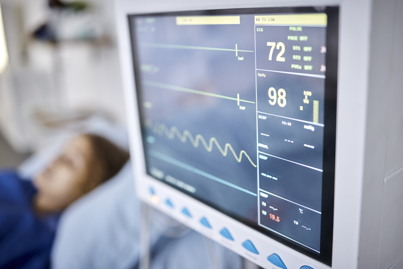
How to Measure Respiratory Rate

To measure respiratory rate or breathing rate, follow these steps:

  • Monitor the person for a few seconds to better understand how they are breathing. The patient should sit or lie but remain still during this period.
  • Count the movements of the chest as it rises or falls. Every time it rises, it indicates one breath.
  • Monitor this rate over 1 minute while watching a clock or a watch.

This indicates the number of breaths per minute.

What Conditions Affect Your Respiratory Rate?

A range of conditions exist that could influence the rate at which you are breathing. Some of the most common include:

  • Chronic obstructive pulmonary disorder (COPD)
  • Anxiety
  • Heart failure
  • Pneumonia
  • Carbon monoxide poisoning
  • Asthma
  • Medications taken
  • Fever or infection
  • Pulmonary embolism
  • Respiratory conditions such as the common cold or flue
  • Dehydration
  • Chronic smoking
  • Pain
  • Anemia

Causes of Abnormal Respiratory Rates in a Child

Common causes of abnormal breathing or respiratory distress in children include the following:

  • Bacterial or viral infections, including croup or pneumonia
  • Allergies
  • Asthma
  • Exposure to cigarette smoke or other harmful gases, including pollution and carbon monoxide
  • A blocked airway (choking)
  • Trauma or damage to any part of the respiratory system
  • Complications from medications (e.g., accidental overdose)
  • Certain genetic conditions, including cystic fibrosis

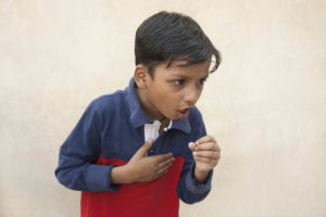
Signs and Symptoms of Pediatric Respiratory Distress

As a general rule, a child who is consistently taking fewer than 10 breaths per minute or greater than 60 breaths per minute is likely suffering from some sort of respiratory distress and requires immediate attention.
Beyond assessing a child’s respiratory rate, you can also look for other key signs and symptoms of pediatric breathing problems. These signs and symptoms include:

    • Changes in skin quality: a child’s skin (especially around the lips, hands, and feet, including the nail beds) might become pale, purplish, grayish, or bluish in color, and could start to feel cold and clammy
    • Nasal flaring: the nostrils open wide when breathing in
    • Chest retraction: with each breath, the chest appears to sink in just below the neck or beneath the breastbone
    • Noisy breathing: a child might begin to make unusual sounds while breathing, including
      • Wheezing (a high-pitched, whistling, musical-like sound that usually only happens on exhalation and usually suggests a narrowing in the lower airway)
      • Stridor (a special kind of high-pitched wheezing noise that can occur during exhalation or inhalation and usually suggests an obstruction or narrowing in the upper airway)
      • Stertor (a snoring-like sound that often develops when a child has congestion in the mouth or nose)
      • Grunting (“ugh” sound)
    • Changes in mood or level of alertness: the child might become drowsy, anxious, restless, difficult to arouse, or simply not like themselves
    • Changes in body position: a child may spontaneously lean forward or tilt their head up or backwards in an attempt to make breathing easier (if a child is choking, he or she may also exhibit the universal sign of choking by placing their hands at their throat)

Untreated, respiratory distress can be fatal or lead to life-threatening or life-altering complications including organ damage, shock, anoxic brain injury, and nervous system depression due to increased carbon dioxide levels in the blood.

Importance of Respiratory Rate in Health

Maintaining the normal respiratory rate for adults and children is critical to health and well-being. Any range outside of the normal respiratory rate indicates that a person’s lungs are not getting enough oxygen to meet the body’s needs. Low respiratory rates often mean the lungs are not breathing in enough air, while a high breathing rate indicates shallow breaths that may not provide enough oxygen. Maintaining normal respiratory rates can greatly enhance the overall function of the brain and other organs.

How to Monitor Your Respiratory Rate

ecg and patient monitor in icuThe only way to monitor your breathing rate manually is to count the number of breaths you have while at rest. Once you learn how to measure your respiratory rate (as noted above), you can track your breathing rate over time to notice differences.

Monitor Respiratory Rates for Children

If you are unsure if your child is getting the normal respiratory rates they need, monitor them to notice changes in breathing rate and function. For example, if your child seems to be breathing heavily or wheezing, that could indicate a lung concern. If you see any of the above-mentioned symptoms, have the child sit down and count their breaths. You can also monitor their respiratory rate during sleep to ensure they are safe. If you do not see normal respiratory rates, consider further investigation to determine if the child is experiencing a health concern.

How to Improve Your Respiratory Rate

If your normal respiratory rates are higher than normal, you can work to improve these rates. To do so, you’ll need to improve overall fitness levels and learn to breathe deeper. In terms of breathing rates, practice makes improvement possible – deep breathing, counting your breaths, and watching your posture can also help.

What Is the Correct Ventilation Rate for a Pediatric Patient in a Respiratory Arrest?

The correct ventilation rate for a pediatric patient is 1 ventilation for every 2 to 3 seconds.

When to Seek Medical Help

When the normal respiratory rates are not met, as noted above by age, it is important to seek medical care. This is especially important when it occurs for more than a few minutes. There is often the need to take corrective action to supply more oxygen to the brain in these situations. A breathing rate while sleeping below the standard levels listed above is indicative of the need for oxygen.

Pediatric vs. Adult Respiratory Rates

The following chart shows normal respiratory rates for children and adults, as well as for newborns, toddlers, and older children.

Respiratory Rates for Children

Age Range Breaths per Minute
Infant (0-12 months) 30-60 breaths per minute
Toddler (1-3 years old) 24-40 breaths per minute
Preschooler (4-5 years) 22-34 breaths per minute
School-aged child (6-12 years old) 18-30 breaths per minute
Adolescence (13-18 years old) 12-16 breaths per minute
Adult 12-18 breaths per minute

What To Do If a Child is In Respiratory Distress (Child vs Infant)

child-coughing
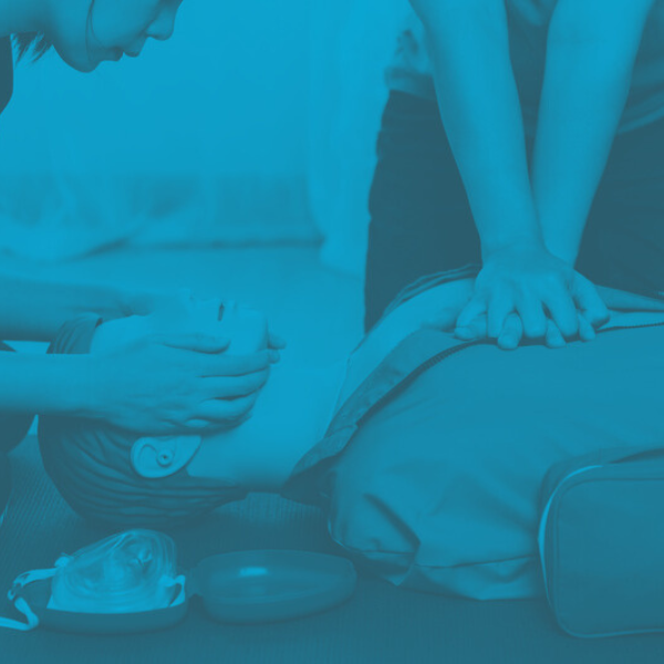
If there is any concern that a child is suffering from respiratory distress, call 911. If a child passes out, begin cardiopulmonary resuscitation (CPR). Importantly, rescuers who are alone should perform CPR on a child for at least 2 minutes before stopping to call 911.

Recall that the CPR techniques used on a baby or prepubescent child (aged 1 to 12 years) have some differences compared to the CPR technique used on an adult. Some of the main differences include the following:

  • For infants, the depth of chest compressions – which can be done with two fingers at the center of the baby’s chest as a single rescuer – should be about one and a half inches (vs two inches for children and adults)
  • Care should be taken not to tip the infant’s or child’s head back too far nor provide rescue breaths that are too forceful
  • Checking a child’s pulse should be performed at the carotid artery (side of the neck), as it is for adults; in infants, the pulse should be checked at the brachial artery (inside of the bicep)

In the case of known or suspected airway obstruction (choking), back blows and/or the abdominal thrust maneuver may be indicated to help the child clear the foreign object.

If a child older than 1 year old is choking, you can provide the same type of abdominal thrust maneuver that would be performed on an adult.

  • First ensure the child is actually choking – look for choking signs and symptoms and ask them, “Are you choking? Can you talk?” If the child is able to speak and cough forcefully, do not begin first aid, as the child may still be able to dislodge the obstruction on their own. If the child is unable to talk or not able to produce a strong and effective cough, proceed with the rescue maneuver.
  • If the child is much shorter, the rescuer should kneel, not stand, behind the choking victim.
  • Wrap your arms around the child’s waist.
  • Place one fist, thumb side in, just above the child’s belly button.
  • Grab your fist with your other hand.
  • Make quick upward and inward movements with your hands, as if you were trying to lift the child up.

For choking children and adults, the American Red Cross recommends using the “five-and-five” approach, in which the rescuer alternates between five black blows and five abdominal thrusts. The rescuer should continue this approach until the object is dislodged, the child begins coughing forcefully again, or the child passes out.

If an infant younger than 1-year-old is choking, a different technique is required (all of these techniques are discussed in detail in BLS training courses and related certifications). To do:

  • Hold the infant face down against your forearm and support the infant’s head and neck securely using your hand. Be sure to keep the infant’s head lower than the chest. If able, support your arm by resting it against your thigh.
  • Use the heel of your other hand to give the child five quick forceful blows between the shoulder blades.
  • Carefully flip the infant over so that she is resting on her back.
    With your other hand, place two fingers on the center of the infant’s chest, just below the nipples. Press your fingers inward quickly five times.

Share your experience of assisting someone in respiratory distress.

Normal Respiratory Rates Are Critical to Know

  • The normal respiratory rate for adults is 12 to 20 per minute and differs for children based on age.
  • A low respiratory rate could mean that a person is not getting the oxygen they need.
  • Knowing the difference in normal respiratory rates for adults and children allows you to know when to take action.

Are Yo​​u Prepared To Care for a Child in Respiratory Distress?

If you’re a healthcare provider or an employer, contact Advanced Medical Certification today to learn about our online certification and recertification courses in Pediatric Advanced Life Support (PALS) and other courses.

About Greta

Greta is a dedicated life saver and a distinguished expert in the field of medical content creation and editing. Her impressive array of certifications in ACLS, CPR, PALS, and BLS underscores her commitment to excellence in the medical field. With over four years of invaluable experience in medical education, Greta plays an indispensable role within the Advanced Medical Certification team, shaping the way healthcare professionals around the world acquire and apply vital knowledge.

Greta's profound expertise serves as the driving force behind the development and distribution of medical content that has significantly enhanced the capabilities of countless healthcare practitioners across the globe.

In addition to her medical qualifications, Greta holds a prestigious academic distinction in Marketing and Global Business from Vilnius University. Her academic journey has been enriched by immersive studies in Slovakia and Portugal during her time as an exchange student, providing her with a global perspective that complements her medical expertise.

Beyond her professional commitments, Greta possesses a genuine passion for global exploration, with a particular focus on immersing herself in diverse cultures and appreciating the intricacies of the natural world. While residing in Vilnius, Lithuania, she continues to make substantial contributions to the field of medical education, leaving an indelible mark on the sector.

Reach out to Greta at greta.kviklyte@advmedcert.com.

Important Things to Know About BLS Renewal

Important Things to Know About BLS Renewal

Photo of Greta

by Greta Kviklyte

Life Saver, AMC
Co-authored by Kim Murray, RN, M.S.

posted on Dec 23, 2021, at 4:13 am

Share

Basic Life Support (BLS) includes a variety of emergency lifesaving techniques and tools—including cardiopulmonary resuscitation (CPR) and automated external defibrillators (AED)—that are offered to people experiencing life-threatening situations, such as an obstructed airway (choking), respiratory distress, and cardiac arrest.

A person who is trained in BLS, such as a healthcare provider or first responder, is expected to have a range of skills as well as the knowledge of how to apply these skills in a variety of situations for people of different ages, including infants, young children, and adults.

In this article, we discuss important things to know about the BLS renewal process, including how often a healthcare provider should renew their certification, common mistakes made during BLS and CPR training courses, and helpful tips to prepare for the BLS renewal course.

BLS Certification Basics: Who Needs It, How to Get It, and How Often It Should Be Renewed

Any healthcare provider, first responder, or medical professional must be certified in BLS. Courses offering BLS for healthcare providers cover a variety of topics, including:

  • How and when to perform CPR for adults, children, and infants
  • How and when to use an AED
  • How to relieve foreign body airway obstruction in adults and infants
  • How to provide effective ventilation using barrier devices
  • Essential life-saving practices, including the Chain of Survival and scene safety
  • The difference between single-rescuer and multi-rescuer during CPR

Many healthcare providers choose or, depending on their professional roles, are required to become trained in additional higher-level certifications such as Advanced Cardiac Life Support (ACLS) and Pediatric Advanced Life Support (PALS).

Do Non-Medical Professionals Need BLS Certification?

practicing-compressions-using-mannequinFor non-medical professionals and laypeople, BLS and other advanced lifesaving certifications aren’t necessary. However, CPR and basic first aid are extremely valuable skills for anyone to know. Individuals who choose to learn CPR and basic first aid could end up saving a life someday—including the life of someone they love or work with.

Consider these important facts from the American Heart Association:

  • Every year, over 350,000 cardiac arrests (heart attacks) occur outside the hospital—most of these (70%) occur in private homes or residences, followed by public settings like parks, malls, and airports (18.8%) and nursing homes (11.2%)
  • According to the Occupational Health and Safety Administration (OHSA), about 10,000 cardiac arrests happen in the workplace every year
  • Effective CPR provided immediately after a person experiences cardiac arrest can double or even triple a person’s chance of survival

Unfortunately, data from the 2017 Heart Disease and Stroke Statistics show that fewer than half (46%) of all people who suffer cardiac arrest outside the hospital ever receive bystander CPR. And according to the U.S. Centers for Disease Control and Prevention (CDC), as many as 9 out of 10 people who have a heart attack outside the hospital die. By encouraging more people to learn how and when to do CPR, these survival odds can improve.

Out-of-Hospital Cardiac Arrest, Women, and CPR

Here’s another interesting fact from the CDC:

Women are less likely than men to receive bystander CPR if they experience cardiac arrest in a public setting. This could be due to many factors:

  • Some people don’t realize women can have heart attacks
  • Women often have atypical warning signs and symptoms of cardiac arrest, including shortness of breath, nausea, vomiting, and back pain or jaw pain
  • Potential rescuers may worry about injuring a woman when giving CPR or being accused of sexual assault
  • People may mistakenly think women are being “dramatic” or “faking it”

Overall, it’s clear that raising awareness about the benefits of CPR and how to provide it is an important public health strategy, especially given the annual incidence of heart attacks—which sadly takes the lives of more people than prostate cancer, flu, pneumonia, car accidents, HIV/AIDS, firearms, and house fires combined.

Do Your Loved Ones Know This About CPR?

  • While formal CPR training can certainly help someone feel more prepared in the event of an emergency, a person does not have to be specially certified to give CPR to someone in cardiac or respiratory distress
  • CPR performed by non-medical professionals and bystanders does not have to involve any breathing into a person’s mouth
  • When performing CPR, people should aim to perform 100 chest compressions per minute—that’s as fast as the beat of the song “Stayin’ Alive”

Where to Receive BLS Training

A variety of organizations provide formal BLS training, including the American Red Cross, the American Heart Association, and Advanced Medical Certification. Healthcare providers may complete their BLS renewal online or in-person, depending on the certifying organization.

To ensure that their BLS certification will be accepted, medical professionals should check with their employer or organization for any specific requirements.

How Often Should A Healthcare Provider Renew Their BLS Certification?

ems-performing-bag-mask-ventilation
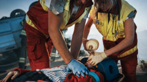
BLS certifications must be renewed every two years. This is to ensure essential lifesaving skills and knowledge are retained, which can help improve outcomes for people suffering from life-threatening emergencies.

It’s also important to consider that failure to maintain an active BLS certification could result in consequences in a healthcare provider’s workplace, including temporary suspension.

Common Mistakes People Make During BLS Certification and BLS Renewal Courses

It’s helpful to be aware of common mistakes people make while providing basic life support, as well as mistakes frequently made by students taking BLS certification or renewal courses. Avoiding these mistakes will ensure that healthcare providers’ skills remain effective and increase the chances of positive outcomes when and if a healthcare provider must provide emergency medical treatment to someone suffering from cardiac arrest, choking, or respiratory distress.

Common BLS mistakes include:

  • Forgetting to ensure scene safety before attending to the patient
  • Forgetting to call 911 immediately upon finding someone in distress
  • Not adequately delegating tasks to other people who can provide assistance (e.g., calling emergency services, finding the AED, etc.)
  • Not utilizing the multiple rescuer technique if it is available (important for reducing rescuer fatigue and ensuring effective lifesaving techniques are used)
  • Not maintaining the correct speed and/or depth of chest compressions
  • Not allowing the chest to fully recoil during CPR prior to performing the next chest compression
  • Not having proper body mechanics while performing CPR (e.g., leaning off to the side of a victim instead of being directly over the victim during chest compressions)
  • Bending the arms during chest compressions
  • If performing rescue breathing, not remembering to open the airway by performing the head tilt/chin lift technique and/or not creating a firm seal around the person’s nose or mouth with the bag valve mask

In addition to errors made while performing lifesaving skills, students should also take care to avoid common mistakes within the course itself which may impede successful course completion. These mistakes include rushing during the test, not carefully reading each question on the test, and not asking the instructor clarifying questions that could otherwise help ensure subject competency.

Quick Reference: How to Perform CPR

These instructions are for single-rescuer medical professionals who have undergone BLS training. Non-medical professionals and bystanders do not need to perform rescue breathing—chest compressions only.

  1. Check scene safety and ensure the person needs help. If available, put on personal protective equipment (PPE) like gloves.
  2. Confirm the victim is unresponsive by using the shout-tap-shout method.
  3. If the person does not respond and isn’t breathing (or only gasping), call 911 and get any available equipment, including an AED, a bag valve mask, and/or a barrier device. If you are not alone, tell someone to call 911 and get these items for you.
  4. Ensure the person is on their back on a firm surface.
  5. Give 30 chest compressions:
  • Two hands on the chest
  • Shoulders directly over hands
  • Elbows straight
  • Compress to at least 2 inches at a rate of 100 to 120 beats per minute
  • Allow the chest to fully recoil (return to its normal position) before performing the next compression

6. Give 2 rescue breaths:

  • Open the airway using a head-tilt/chin-lift technique
  • Give each rescue breath for one second, watching to make sure the chest rises (if the chest doesn’t rise, tilt the head and ensure a proper seal before trying a second breath—there could be an airway obstruction if the chest still doesn’t rise)

7. Continue giving 30 chest compressions and 2 breaths until help or an AED arrives.

Quick Reference: How to Perform the Abdominal Thrust Technique

The abdominal thrust technique is to be used when a non-pregnant adult or child over age one is choking. Before initiating the abdominal thrust technique, ensure the person is choking by observing for signs and symptoms (e.g., the universal sign of choking/hands at the throat, noisy or difficulty breathing, inability to talk, laugh, or cry, weak or ineffective cough, flushed skin that could be bluish or pale; you can also ask, “Are you choking?” and look to see if the person nods).

    1. Tell the person you are going to help.
    2. Stand behind the person with your knees slightly bent and your feet staggered to improve your balance. If it is a child, kneel behind them.
    3. Wrap your arms around the person’s waist.
    4. Make a fist with one hand and put it slightly above the person’s belly button.
    5. Grasp your fist with your other hand.
    6. Press hard into the person’s abdomen with a quick upward movement.
    7. Perform 6 to 10 abdominal thrusts until the object is dislodged.
    8. If the person passes out, begin CPR.

Note: the American Red Cross recommends a “five and five” technique by alternating between five back blows and five abdominal thrusts until the object blocking the airway is dislodged, or until the person passes out.

Quick Reference: How to Use an Automated External Defibrillator (AED)

AEDs are intended for non-breathing adults and children aged 8 or older who weigh at least 55 pounds.

    1. Check scene safety and ensure the person needs help.
    2. If you’re not alone, ask a bystander to call 911. If you don’t have an AED, ask a bystander to get one for you.
    3. Initiate CPR until the AED becomes available.
    4. As soon as an AED becomes available, turn it on and follow the prompts. If you’re not alone, one person can continue performing CPR while the other person gets the AED ready.
    5. Remove all clothing covering the victim’s chest. If necessary, wipe the chest dry.
    6. Attach the AED pads as instructed, depending on whether the victim is an adult or a child. The pad packets will have diagrams to show you how.
    7. Make sure the pad connector cable is connected to the AED.
    8. Prepare to allow the AED to analyze the victim’s heart rhythm by making sure no one is touching the victim—say “CLEAR!” in a loud and commanding voice.
    9. If the AED determines a shock is needed, make sure no one is touching the victim (say “CLEAR!” in a loud and commanding voice) and press the “shock” button to deliver the shock.
    10. After the shock is delivered and the AED determines no shock is advised, immediately restart giving CPR, starting with chest compressions

Preparing For Your BLS Renewal: 3 Tips for Success

1. Get prepared to get hands-on. Many of the skills learned in BLS courses involve physical techniques such as the abdominal thrust maneuver and CPR. Practicing these skills is important for helping you prepare for real-life emergencies, where fast, accurate action is imperative.
Be sure to practice your physical skills even if you are taking your BLS certification course completely online.

2. If you’re not sure about something, ask. Developing mastery and familiarity with BLS subject matter can make the difference between life and death. Students should never hesitate to ask their BLS course instructor questions about the course material. Chances are, many people will have similar questions and would benefit from further clarification.

3. Choose a reputable organization for your BLS certification course. You have many choices when it comes to BLS certification and renewals, so be sure to select a company known for offering high-quality training and support.

In an effort to provide quality, cost-effective resources for our students, Advanced Medical Certification course materials are based on the latest best practice resources, including the International Liaison Committee on Resuscitation (ILCOR) 2020-2025 Edition ACLS Provider Handbook. Our courses are also eligible for continuing medical education (CME) credits and can be completed in the convenience and privacy of one’s own home or workplace.

Our website is one of fewer than 1% of sites on the internet endorsed by HONCode, a UN chartered non-governmental agency that seeks to ensure quality health information is shared for patients, providers, and the public.

AMC proudly features a 98% national acceptance rate. In the unlikely scenario that your employer does not accept our certification, we’re happy to offer a full refund.

Conclusion

If you’re a healthcare provider, first responder, or medical student, it’s important to ensure your BLS certification—and your basic lifesaving skills—are up-to-date. To begin comprehensive and cost-effective training for yourself or your workforce, or to learn more about BLS recertification online, contact Advanced Medical Certification today. Share a story of starting your BLS certification.

About Greta

Greta is a dedicated life saver and a distinguished expert in the field of medical content creation and editing. Her impressive array of certifications in ACLS, CPR, PALS, and BLS underscores her commitment to excellence in the medical field. With over four years of invaluable experience in medical education, Greta plays an indispensable role within the Advanced Medical Certification team, shaping the way healthcare professionals around the world acquire and apply vital knowledge.

Greta's profound expertise serves as the driving force behind the development and distribution of medical content that has significantly enhanced the capabilities of countless healthcare practitioners across the globe.

In addition to her medical qualifications, Greta holds a prestigious academic distinction in Marketing and Global Business from Vilnius University. Her academic journey has been enriched by immersive studies in Slovakia and Portugal during her time as an exchange student, providing her with a global perspective that complements her medical expertise.

Beyond her professional commitments, Greta possesses a genuine passion for global exploration, with a particular focus on immersing herself in diverse cultures and appreciating the intricacies of the natural world. While residing in Vilnius, Lithuania, she continues to make substantial contributions to the field of medical education, leaving an indelible mark on the sector.

Reach out to Greta at greta.kviklyte@advmedcert.com.

Vagal Maneuvers Made Simple: Combat SVT Without Meds

Vagal Maneuvers Made Simple: Combat SVT Without Meds

Photo of Greta

by Greta Kviklyte

Life Saver, AMC
Co-authored by Kim Murray, RN, M.S.

posted on Nov 27, 2021, at 4:42 am

Share

For those suffering from supraventricular tachycardia, it may be possible to find improvement through the use of vagal maneuvers. Find out what they are and what this type of treatment could mean for you.

What Are Vagal Maneuvers?

Vagal maneuvers are a type of physical action that you can take to make the vagus nerve slow down its electrical impulses. The vagus nerve, which is the heart’s natural pacemaker, runs from the brainstem to your midsection. It plays a role in your heart rate.

What Is SVT?

Supraventricular tachycardia (SVT), sometimes called paroxysmal supraventricular tachycardia, refers to a group of conditions affecting the upper chambers of the heart (atria). These conditions occur due to faulty electrical signaling within the heart and cause the heart to beat unusually fast, for as little as a few seconds to as much as a few hours or days.

SVT is rarely fatal, but it can lead to signs and symptoms that are uncomfortable and distressing, including chest pain, shortness of breath, sweating, lightheadedness, dizziness, fainting (syncope) or near fainting, fatigue, pulsing sensations in the neck, and heart palpitations (the feeling that the heart is fluttering, pounding, murmuring, throbbing, or skipping a beat).

To help people manage SVT, doctors rely on multiple interventions, including medications, procedures like ablation or electrocardioversion, and vagal maneuvers. One benefit of vagal maneuvers is that patients can learn how to perform these maneuvers on their own, which helps them manage their supraventricular tachycardia symptoms at home.

This blog post discusses what vagal maneuvers are, how they are used to help slow down a person’s heartbeat, and what safety considerations patients and medical providers should be aware of before using vagal maneuvers as part of the first-line treatment for tachycardia.

Did You Know?

old man consulting a doctorThe normal resting heartbeat of an adult is between 60 and 100 beats per minute. The normal resting heart rate of an infant (birth to about 3 months of age) ranges from
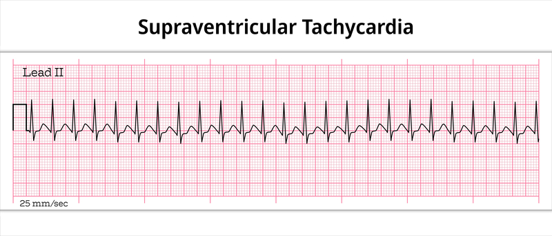
 100 to 150 beats per minute. Between one and three years of age, a child’s resting heart rate should fall between 70 and 110 beats per minute.

By the time a child reaches age 12, their resting heart rate more closely resembles an adult’s, ranging from 55 to 85 beats per minute.

SVT is the most common kind of abnormal heart rhythm in children. But while abnormal heart rhythms can lead to cardiac events in children and infants, SVT is almost never life-threatening in kids or adults.

Understanding Vagal Maneuvers and Their Role in SVT

The vagus nerve (also known as the tenth cranial nerve or CN X) is an important part of the body’s parasympathetic nervous system, which is the “rest and digest” part of the nervous system that facilitates relaxation. The vagus nerve—which travels from the brainstem to the abdomen—helps control things like mood, digestion, and immune function. It also innervates heart muscle fibers and helps control the heart rate.

Vagal maneuvers are techniques intended to stimulate the vagus nerve, which can help slow down electrical activity in a structure in the heart called the atrioventricular (AV) node. To understand why vagal nerve activation is important for managing supraventricular tachycardia, it’s helpful to know a little more about the different types of SVT.

The most common type of SVT is called atrioventricular nodal reentrant tachycardia (AVNRT). AVRNT occurs when there is an extra electrical pathway (reentrant circuit) in or near the AV node that causes the heart to beat too fast.

Another type of SVT is called atrial tachycardia, which includes atrial flutter (the upper chambers of the heartbeat faster than the lower chambers) and atrial fibrillation (the upper chambers of the heartbeat fast and irregularly).

Common risk factors for SVT include family history, heavy smoking or alcohol use, excess caffeine, intense exercise, high stress, drug use, certain medications, pregnancy, advancing age, and underlying medical conditions like lung problems, obesity, thyroid diseases, or other heart valve diseases.

According to 2015 clinical guidelines from the American Heart Association, American College of Cardiology, and Heart Rhythm Society, SVT is common in both outpatient clinical practice and emergency medicine settings. These 2015 clinical guidelines also recommend vagal maneuvers as one of the first lines of treatment for people with SVT.

Types of Vagal Maneuvers

The following are the most common vagal maneuvers recommended by healthcare providers at The Cleveland Clinic

  • Valsalva maneuver: Also known as bearing down like you are going to have a bowel movement
  • Diving reflex
  • Gag reflex
  • Carotid sinus massage
  • Coughing
  • Handstands for 30 seconds
  • Applied abdominal pressure

If you want to stop SVT attacks, using one of these recommended vagal maneuvers for SVT can make a big difference.

Vagal Maneuvers in Children

It is possible to learn how to stop SVT attacks with vagal maneuvers in children. Steps may include the following. Have the child calm down and lie down for five minutes. If the heart rate remains elevated, it may be SVT. If that occurs, choose one of the vagal maneuvers to apply:

  • Valsalva maneuver
  • Turn the child upside down by their ankles or have the child do a handstand with support
  • A finger or popsicle placed at the back of the throat to stimulate a gag reflex
  • Facial ice water immersion can also help

5 Vagal Maneuvers to Manage Supraventricular Tachycardia

Supraventricular Tachycardia - Electrocardiography PaperIn a 2014 review, Sohinki and Obel note that vagal maneuvers are effective at reversing SVT and returning the heart to a normal rate (sinus rhythm) about 25% of the time, on average. These maneuvers can be performed in a variety of ways. Five techniques are described here:

1. Valsalva Maneuver (Bearing Down)

Many people perform Valsalva maneuvers in daily life, such as when they are lifting something heavy or straining to defecate. Generally speaking, this technique involves a forceful attempt to breathe out even though the airway is closed.

To perform a Valsalva maneuver for SVT in the clinical setting, a patient should lay down in a semi-recumbent (partially reclined) position. They should forcefully exhale against resistance into a pressure measuring device called a manometer (or, alternatively, a large syringe) for about 15 seconds, generating a pressure of 40 mmHg.

Next, the patient lays flat and the provider passively raises the patient’s legs to about 45 degrees. After 15 seconds, the patient returns to the semi-recumbent position. During this maneuver, the patient’s heart rate, blood pressure, and other vital signs are monitored closely.

The Valsalva maneuver is expected to create a physiological response that can be broken down into four phases:

  • Phase 1: forced exhalation against resistance increases pressure inside the chest, which temporarily increases pressure inside the aorta (the main artery that carries blood away from the heart); to compensate for this increased aortic pressure, the heart rate decreases
  • Phase 2: the increased pressure inside the chest temporarily reduces the amount of blood flowing back to the heart (venous return) and therefore reduces the amount of blood pumped out by the heart (cardiac output); to compensate, the heart rate starts to increase again
  • Phase 3: at the end of the forceful exhalation, the pressure inside the aorta decreases, further leading to a compensatory rise in heart rate
  • Phase 4: having the patient lay flat with their legs raised increases the venous return to the heart, which again increases the aortic pressure and leads to a compensatory decrease in heart rate

If the Valsalva maneuver works correctly, a person’s heart should return to its normal resting rate once all four phases are complete.

2. Coughing

The Valsalva maneuver has a similar effect to sustained forceful coughing. One benefit of coughing is that it is usually easier for patients to do.

3. Cold Stimulus to the Face

The diver’s reflex, recognized as early as the 1700s, is a physiological reflex in humans and other mammals that is triggered in response to holding the breath while submerged in cold water. This causes the heart rate to decrease and the pressure inside peripheral veins to increase, which decreases the amount of work the heart has to do and therefore helps conserve oxygen.

People experiencing SVT can simulate their diver’s reflex by submerging their face in a container of ice or ice-cold water or putting an ice-cold washcloth or icepack on their face. Each type of cold stimulus should last about 10 seconds.

4. Carotid Massage

Only a trained medical professional such as a doctor should perform a carotid massage, which is done while the patient’s neck is extended back and turned away from the side being massaged. This maneuver may help treat SVT by stimulating nerve endings in the carotid arteries that tell the heart to slow down.

To perform this technique, a doctor moves his or her fingers in a gentle circular motion over the carotid artery just beneath the jaw for about 10 seconds. Only one side of the neck should be massaged at a time.

Carotid massage can be used to both diagnose and treat SVT.

5. Gagging

The gagging reflex

Gagging stimulates the vagus nerve and can stop an episode of SVT. A tongue depressor is briefly inserted into the patient’s mouth, touching the back of the throat, which causes the person to reflexively gag. The gag reflex stimulates the vagus nerve.

Why Are Vagal Maneuvers Used?

Numerous factors relate to the benefits of using vagal maneuvers. The goal of performing any of these methods is to slow down the rate the heart is beating, ultimately overcoming an SVT attack.

Who Shouldn’t Have Vagal Maneuvers?

There are some situations in which a person should not engage in any of these methods. Including those who have:

  • Low blood pressure
  • Chest pain
  • Shortness of breath
  • Severe valvular disease
  • Recent strokes
  • Uncontrolled high blood pressure

How Commonly Are Vagal Maneuvers Used?

SVT is a common condition that occurs in children and adults. Data from the Cleveland Clinic indicates that an estimated 1 in 250 people to 1 in 1000 children have SVT. Vagal maneuvers are the first line of treatment for this condition. That makes them very commonly used.

What Happens Before Vagal Maneuvers?

In a hospital setting, a provider will complete an electrocardiogram to better understand the heart’s rate of beating. They will also monitor blood pressure and heart rate while keeping a close eye on your oxygen rates. By understanding these factors, rescuers can determine if the patient is struggling with anxiety or intense physical stress or if they are experiencing an SVT attack.

What Happens After Vagal Maneuvers?

In situations where vagal maneuvers are used, it may be necessary to follow up to determine if the underlying cause of the condition can be estimated. This may include completing an ECG or ECHO to learn more about the structure of the heart. Often, a person will need some time to rest after treatment, but no additional treatments or steps are necessary if SVT ends.

What Are the Advantages of Vagal Maneuvers?

The core benefit of using the supraventricular tachycardia vagal maneuver is that it is a non-invasive way of naturally helping your body reset its cardiac rhythm. It does not involve any type of medication or advanced treatment, and it can be very effective for many patients.

What Are the Risks of Complications of Vagal Maneuvers?

healthcare professionals performing cpr
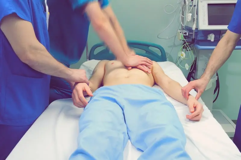
While you may wish to learn how to lower your heart rate using a supraventricular tachycardia vagal maneuver, some risks go along with it.

  • Hypotension, low blood pressure
  • Bradycardia, a slow heart rate
  • Ventricular tachycardia, very rate
  • Ventricular fibrillation has, a very high rate
  • Atrial fibrillation is very rare

Recovery and Outlook

In an estimated 20% to 40% of patients, the SVT will improve. This is one of the most effective ways to lower your heart rate. However, it can happen again, and that may mean it is necessary to consider medication to improve your heart rate or more advanced treatments such as ablation.

What Is the Outlook for Vagal Maneuvers?

The use of vagal maneuvers can lead to positive outcomes for many people. Most people do not experience any complications from this procedure, making it very safe.

When to Call the Doctor

If you are asking, “What is the Valsalva maneuver?” or you want to know when to apply this treatment, call your doctor. If lying down does not calm your heart rate, then call your doctor. You can learn how to stop SVT attacks using these methods, but if you continue to experience them, it is critical to seek care.

Precautions and Contraindications

Patient having a cardiac attackWhile they are simple, non-invasive, and generally effective, vagal maneuvers are not without risk. Patients should never attempt vagal maneuvers without the supervision of a medical provider.

For example, carotid massage is contraindicated in anyone who has a history of ventricular fibrillation or ventricular tachycardia, or in anyone who has had a heart attack, stroke, or transient ischemic attack (TIA or mini-stroke) less than three months ago.

Valsalva maneuvers are contraindicated in patients who have SVT along with:

  • An acute heart attack
  • Unstable blood pressure (e.g., systolic blood pressure less than 90 mmHg)
  • Aortic stenosis
  • Carotid artery stenosis
  • Certain eye disorders, including glaucoma or retinopathy

Vagal maneuvers may also lead to adverse events including, syncope or heart block (when the electrical signals to the heart are blocked).

One 2018 case report by Nakamori et al described the case of a 79-year-old male who developed atrial fibrillation after performing the Valsalva maneuver when he had SVT following heart surgery. Atrial fibrillation is widely considered the most dangerous type of SVT since it can lead to potentially fatal complications including heart failure, blood clots, and stroke. In this particular case, doctors used medication to help restore the patient’s normal heart rate, which returned within four hours.

Other Treatment and Prevention Options for SVT

As noted, current research suggests that the effectiveness of vagal maneuvers to treat SVT is about 25%, with a success rate ranging from as low as 6% to as high as 54%, depending on the study. What this suggests is that other interventions are often necessary to help terminate tachycardia and reestablish a normal heart rhythm.

These interventions may include:

  • Cardioversion, in which an electric current is delivered to the heart via patches or paddles on the chest, which helps “shock” the heart back into a normal rhythm
  • Ablation, in which a doctor inserts a thin flexible tube called a catheter through a vein or artery (usually in the groin), then uses cold or heat sensors on the end of the catheter to create tiny scars in certain parts of the heart to block faulty electrical signals

In rare instances, some people who experience SVT are treated with a pacemaker. A pacemaker is a small device implanted beneath the skin near the collarbone that helps control a person’s heartbeat.

Individuals can also reduce their risk of SVT and prevent future episodes by making simple and healthy lifestyle changes. This includes:

    • Understanding what causes tachycardia for them (identifying personal triggers)
    • Not smoking
    • Minimizing or eliminating alcohol and caffeine
    • Managing stress levels
    • Getting enough sleep
  • Maintaining a healthy weight (a 2018 study published in The American Journal of Cardiology with more than 67,000 participants found that people who are obese were 40% more likely to develop atrial fibrillation than people who weren’t obese)
  • Using medications only as prescribed and knowing which kinds of medications can lead to a rapid heart rate, including antidepressants, blood pressure medications, and even over-the-counter decongestants or cold medications
  • Avoiding illegal drugs, including cocaine, ecstasy, or methamphetamines
  • Managing other underlying health conditions with the help of a medical provider

Not only can these healthy lifestyle choices help reduce the risk of SVT, but they can also improve overall cardiovascular health.

Conclusion

SVT includes a group of conditions that can cause an abnormally fast heartbeat that can come and go or persist for hours or days. Associated SVT symptoms like lightheadedness, fatigue, and shortness of breath can be incredibly distressing—and can be caused by other health conditions, too. So, always speak with a doctor to get a proper diagnosis.

It’s important to remember that SVT is usually not life-threatening on its own, but certain subtypes of SVT (e.g., atrial fibrillation) may increase the risk of potentially fatal conditions like blood clots, heart attack, or stroke. To learn skills that could save a life, or to update or renew a CPR or First Aid certification conveniently and completely online check Advanced Medical Certification. Share your story of how CPR or First Aid training helped you.

About Greta

Greta is a dedicated life saver and a distinguished expert in the field of medical content creation and editing. Her impressive array of certifications in ACLS, CPR, PALS, and BLS underscores her commitment to excellence in the medical field. With over four years of invaluable experience in medical education, Greta plays an indispensable role within the Advanced Medical Certification team, shaping the way healthcare professionals around the world acquire and apply vital knowledge.

Greta's profound expertise serves as the driving force behind the development and distribution of medical content that has significantly enhanced the capabilities of countless healthcare practitioners across the globe.

In addition to her medical qualifications, Greta holds a prestigious academic distinction in Marketing and Global Business from Vilnius University. Her academic journey has been enriched by immersive studies in Slovakia and Portugal during her time as an exchange student, providing her with a global perspective that complements her medical expertise.

Beyond her professional commitments, Greta possesses a genuine passion for global exploration, with a particular focus on immersing herself in diverse cultures and appreciating the intricacies of the natural world. While residing in Vilnius, Lithuania, she continues to make substantial contributions to the field of medical education, leaving an indelible mark on the sector.

Reach out to Greta at greta.kviklyte@advmedcert.com.

Which Certifications are Required to Become a Medical Assistant?

Which Certifications are Required to Become a Medical Assistant?

Photo of Greta

by Greta Kviklyte

Life Saver, AMC
Co-authored by Kim Murray, RN, M.S.

posted on Nov 23, 2021, at 2:32 am

Share

So, you’re interested in working in health facilities with doctors, nurses, patients, and other health care professionals, but don’t want to attend medical school because it’s time-consuming and expensive. Whatever the reason, consider becoming a medical assistant.

Medical assistants have no certification requirements, but there are different types of education, certifications, licensure, and training that are often recommended or offered by specific health care facilities. These can influence your professional career trajectory.

Read on to explore the education and certification requirements for this professional career path.

What Is a Medical Assistant?

female-surgeonMedical assistants usually take medical histories and vitals from patients. They may assist in more advanced ways like drawing blood and providing first aid as their skills advance. You may also handle important administrative, clerical and clinical tasks as you learn more on the job. You might work in a doctor’s office, community clinic, hospital, or another clinical setting.

A medical assistant must have specific personality and character traits as well as the skills needed for the job. First of all, you need to have a desire and passion to help, listen and guide any patient needing help.

A typical day involves moving quickly on your feet, solving various problems, practicing good communication among faculty, and keeping on your toes. You’re expected to perform a mix of administrative, clinical, and clerical tasks as needed by the employer, so your role may change from day to day.

This aspect of the job can keep medical assistance interesting. You’re often learning something new and adding to your responsibilities. That’s all good for advancing your career.

Listing every possible job duty of a medical assistant would be lengthy considering their valued and crucial position in the office. Above all, your roles will always be centered on the needs of the patient and the requests of the physician.

Is This a Good Career Path?

It can be for many people. Medical Assistants are in great demand. This professional career is growing 18% over the next 10 years, which is much faster than others.

According to the Bureau of Labor Statistics (BLS.GOV), the US median salary for a medical assistant is around $17/hr plus good benefits as of 2020. Although it can vary a lot from state to state. In Washington, for example, $27/hr is more common but the cost of living is also higher.

This pay can vary based on education level, certification, experience, and job performance.

How Do You Become a Medical Assistant?

The answer to this question varies. But there are numerous steps you can take to improve employment opportunities.

On the one hand, most states don’t require you to have any formal education after high school to become a medical assistant. You do have to have graduated from high school or gotten your GED.

Many medical assistants become one by taking a position that requires no experience. They learn on the job. They then take on more responsibilities as they learn more. Once you’ve been working in this job for a few years, you might decide to become certified.

However, like many medical careers right now, things are changing for Medical Assistants.

Many employers now look for people who have completed more advanced training which can take one to two years.

According to a 2020 report conducted by the National Healthcare Association. 89% of employers now encourage their medical assistants to get certified if they aren’t already. 63% pay more to Certified Medical Assistants solely based on their professional certification.

Because your level of education and experience directly impact your pay, you’ll likely want to explore additional certifications to further your career. Note: The Bureau of Labor Statistics based its median pay rate above on the assumption that you are certified.

What Education Does a Medical Assistant Need?

There are essentially three types of voluntary medical assistant certification:

  • Registered Medical Assistant (RMA)
  • Certified Medical Assistant (CMA)
  • Certified Clinical Medical Assistant (CCMA)

These are offered by different certifying organizations. But they mostly consist of similar curriculum like anatomy and physiology, medical terminology, and other topics including first aid.

For example, the American Association of Medical Assistants offers a nationally recognized Commission on Accreditation of Allied Health Education Programs (CAAHEP) or Accrediting Bureau of Health Education Schools (ABHES) accredited programs to better prepare prospective medical assistants for their career.

Any graduate of the CAAHEP and ABHES programs is eligible to take the certification exam, which covers the program’s material.

Some of this content includes:

  • Pathology
  • Medical terminology
  • Keyboarding and computer applications
  • Record keeping
  • Coding

Aside from the program, you’ll complete a practicum. This is common in medical education and involves a hands-on application to show you can do what you’ve been learning in classwork in real life.

Your certification program may also require an internship or a number of years working as a medical assistant before you can become certified. For example, a potential RMA must have five years of experience as a medical assistant before applying to become an RMA.

Medical Assistant Vs. Certified Medical Assistant

healthcare-professionals-in-the-operating-roomWhile there are no certifications required to become a medical assistant, the material you learn in programs will be extremely beneficial to you and your potential employers. Certification helps you practice to the highest degree, whether you are training to become a medical assistant or a surgeon!

Generally, a Certified Medical Assistant has graduated from an accredited program and taken and passed the certification test.

According to the American Association of Medical Assistants (AAMA-NTL.org), a graduate of a program must take and pass the test within three years of completing their education. A CMA is good for 60 months (five years) and is renewed by re-taking an updated recertification test.

According to the AAMA website, you need a combination of administrative, clinical, and general credits, so be sure to check on the exact requirements of your certifying organizations.

CMA Vs. CCMA Vs. CMAA Vs. RMA

As a Medical Assistant, it’s essential to know what all these letters mean. It may come up in your job search and career.
The American Association of Medical Assistants isn’t the only organization that certifies medical assistants. That’s why the CMA isn’t the only certificate you might encounter.

The National Healthcare Association (NHA) and the American Medical Technologists (AMT) also certify medical assistants. A Registered Medical Assistant (RMA) obtains certification through AMT. A Certified Clinical Medical Assistant (CCMA) obtains certification through NHA.

The RMA, CCMA, and CMA will be considered interchangeable by most employers.

Another certificate through NHA is the CMAA. This is not a medical assistant certificate but could be confused with one.

A CCMA is a Certified Clinical Medical Assistant. Hence, their education focuses more heavily on the clinical side of being a medical assistant, but they can do everything a CMA or RMA can do. This is a medical assistant.

Alternatively, a CMAA is a Certified Medical Administrative Assistant. Their education focuses only on the administrative side, and they are not considered medical assistants despite having “medical” in the name.

This difference is reflected in the education requirement. You can complete a CMAA program in half the time because you’re only focusing on the administrative side of medical assistance. With that said, a CMAA can always learn the medical side on the job to expand their role in states that allow that.

How to Get Recertified

Recertification requirements are slightly different from the CMA vs. the CCMA.

CMA Recertification Process

Before taking the recertification test, the Certified Medical Assistant must acquire 60 recertification points (credits). You obtain these points by completing Continuing Education (CE) relevant to being a medical assistant. CE is also sometimes called CEU Continuing Education Units and CME Continuing Medical Education.

Sixty may sound like a lot. But keep in mind, most CEU courses will give you up to six credits and may only take 4-12 hours to complete. On top of that, these days, you can meet most of them 100% online in self-paced courses. Plus, you get to meet them over five years.

This continuing education requirement is typical for any certified professional. It ensures you’re staying current on best practices and procedures related to your scope of practice.

CCMA Recertification

The CCMA must renew every two years but are only required to obtain 10 CEU credits for their renewals. Like CMA recertification, you can take internal courses through the certifying organization, usually for a per-course fee, or complete outside courses and submit the certificate to them.

What If You Let Your Certification Lapse?

It would help if you always tried to complete all of your requirements well in advance of the deadline to avoid a lapse in your certificate. If it does lapse, you’ll owe a reinstatement fee, which is typically around $100 on top of your recertification fee, which runs around $200-300 dollars.

Word of caution: If you let your certificate lapse for more than 12 months, you may have to re-take certain courses and the original certification exam, which is a lot harder and more costly than the recertification process.

How to Obtain CEU the Easy Way

When you first get certified, two years or five years seems so far away. But it will quickly sneak up on you. So you need a plan to get as many CEU as possible as early as possible.

Taking online advanced medical courses to get medical certifications is one of the best ways to do this. Not only will these prepare you for seamless recertification. They provide you with invaluable medical education that you can use now to show your employer that you’re committed to learning more and doing the best job. This may also lead to additional responsibilities within the scope of medical assistance.

You can leverage this to further your career.

Blood Borne Pathogens Certification

As a medical assistant, you’re likely to be interacting with bodily fluids, including blood, bacteria, viruses, etc. It’s important that you know how to work with these possible contagions in a safe, sanitary, and OSHA-compliant way. In a bloodborne pathogens certification course, you learn how to do that.

This 100% online certification course can provide you with three credits toward your recertification as a Medical Assistant. In the course, you learn practical, real-world ways to work and prepare, store, and manage substances that may contain pathogens.

Even if you’re not currently up for recertification or have yet to become a Certified Medical Assistant, getting a Bloodborne Pathogens certificate looks great on your resume. Medical employers want to know that the people they’re hiring take the possible spread of disease seriously and know how to contain those risks.

CPR, AED, and First Aid Certification

Another great certificate for a Medical Assistant is CPR, AED, and First Aid. First of all, you’ll learn a lot of practical skills that Medical Assistants perform regularly, like maintaining a clean environment, checking vitals, assessing patient distress, managing trauma, and providing first aid for a variety of medical emergencies.

You’ll also learn how to properly perform CPR in a life or death situation and use an Automatic External Defibrillator (AED) to shock the heart back to a normal rhythm. This certification earns up to six CEU to put toward your recertification. They also look great on a resume.

Basic Life Support

Getting certified in BLS is the step up from learning bystander CPR and first aid. This course is designed for medical professionals like you who may need to assist doctors and nurses during a cardiac arrest or similar life-threatening event.

It’s important to mention that getting certified in BLS doesn’t mean you can perform medical procedures that are beyond what a medical assistant does. However, you’ll learn how doctors and nurses handle these events and better understand how you can assist during these high-pressure situations.

A BLS certification counts toward four CEU credits.

Other Advanced Medical Certifications

By far, the above three are the most important for a Medical Assistant. But you can certainly take more advanced courses for medical certifications. If you really want to challenge yourself, consider Advanced Cardiac Life Support (ACLS) and Pediatric Advanced Cardiac Life Support (PALS).

These courses will be more relevant to a registered nurse, doctor, or EMT because they describe advanced medical procedures. But once you’ve completed the others and want to take your understanding to the next level, these provide a more in-depth look at how to manage a life-threatening cardio-respiratory event.

Your Career Advanced

Medical assistance is a rewarding professional career. Like many career paths, obtaining additional certifications can help you advance and meet your life goals. Most importantly, you never want to stop learning because, the more you know, the better you’ll be able to support the patients you work with. Share with us your experience of becoming a medical assistant.

About Greta

Greta is a dedicated life saver and a distinguished expert in the field of medical content creation and editing. Her impressive array of certifications in ACLS, CPR, PALS, and BLS underscores her commitment to excellence in the medical field. With over four years of invaluable experience in medical education, Greta plays an indispensable role within the Advanced Medical Certification team, shaping the way healthcare professionals around the world acquire and apply vital knowledge.

Greta's profound expertise serves as the driving force behind the development and distribution of medical content that has significantly enhanced the capabilities of countless healthcare practitioners across the globe.

In addition to her medical qualifications, Greta holds a prestigious academic distinction in Marketing and Global Business from Vilnius University. Her academic journey has been enriched by immersive studies in Slovakia and Portugal during her time as an exchange student, providing her with a global perspective that complements her medical expertise.

Beyond her professional commitments, Greta possesses a genuine passion for global exploration, with a particular focus on immersing herself in diverse cultures and appreciating the intricacies of the natural world. While residing in Vilnius, Lithuania, she continues to make substantial contributions to the field of medical education, leaving an indelible mark on the sector.

Reach out to Greta at greta.kviklyte@advmedcert.com.

Can You Do CPR Without Formal CPR Training?

Can You Do CPR Without Formal CPR Training?

Photo of Greta

by Greta Kviklyte

Life Saver, AMC
Co-authored by Kim Murray, RN, M.S.

posted on Oct 27, 2021, at 5:52 am

Share

Did you know that 90% of people who have a cardiac arrest while not at the hospital will die (CDC.gov)? That’s a sobering statistic but also one that we shouldn’t ignore.

Yet, here is an even more important fact to know. When someone in the room knows CPR, it increases an individual’s chance of survival by 100 to 200%. That’s huge.

But people are more than numbers. When that individual person is your high school student, parent, or workplace friend, that increased survival rate means everything to you and to the many other people who love them.

CPR is a vital skill like driving, cooking your own food, or performing first aid to control bleeding. Everyone should learn it. But do you need formal CPR training to do it? Can only CPR-certified people perform CPR?

Read on to find out.

Do You Need a CPR Certification to Perform CPR?

The short answer is “no”. You don’t have to be CPR certified to perform CPR.

The purpose of a CPR certificate is to verify that you went through CPR training and took a certification test to show that you retained the skills you learned.

Like so many things in life, you do not have to have a certificate from someone else to be able to do CPR. You’re unlikely to be fined, sued, or suffer any other penalties.

Most states have Good Samaritan laws that protect people legally when they try to help someone, even if the result doesn’t turn out as they expected. And that’s important because every year, around 350,000 cardiac arrests happen at a time when bystander CPR may be the difference between life and death.

According to the Centers for Disease Control and Prevention, 70% of those who have this heart event will experience it in their homes.

What to Do If Someone Needs CPR

Even if you do not have CPR training, if you believe someone has a cardiac arrest, you should:

  • Call 911 and call out for help
  • Ask someone to bring an Automated External Defibrillator (AED). Almost every workplace and school will have one.
  • Start chest compressions. Let the chest come back up in between compressions. The goal is 100-120 compressions per minute. To keep rhythm, use popular songs like “Eye of the Tiger” from Rocky III, Gloria Gaynor’s “I Will Survive”, or Britney Spears’ “Stronger”.
  • Do not stop CPR until emergency help arrives.
  • If someone knows how to use an AED, they should turn it on and follow the verbal instructions.

Note: This is an abbreviated version of CPR training and only explains what someone who isn’t trained could do. And you may be successful.

But there are many reasons formal CPR training is better.

Why Is CPR Training Important?

classroom-based-cpr-trainingIt’s not just CPR that saves lives. It’s knowing how to do it correctly. Someone with no formal CPR training could use what they’ve seen on TV to keep some blood flowing. But they may not be able to do it in a way that improves that person’s chance of survival. It’s unlikely you could perform high-quality compressions without some formal training.

Beyond this obvious point, there are some more subtle reasons to take the time and get formal training.

More Confidence = Better Performance

Studies such as this one published on ScienceDaily.com show that as confidence rises, so does a person’s ability to perform a task effectively.

The relationship between the two is obviously not one way. When you know you know how to do something, you will do it better with less hesitation, leading to better outcomes. At the same time, the CPR training you completed gave you that confidence you needed to perform well.

Obtaining Proficiency

There is a clear difference between knowing how to do something and doing it well. When you complete a CPR training course in the high school gym, workplace, or online, those courses are based upon scientifically proven best practices.

Completing and updating your CPR training every two years ensures you are using the most current evidence-based methods.

Knowing How to Use the Equipment

CPR training teaches you how to use an Automated External Defibrillator (AED). This medical device is not only reserved for medical professionals. Anyone can use it with some basic training.

It delivers electrical shocks to the heart to restore a normal rhythm. But there is a right time to use an AED and the proper way to use it. You learn about this during CPR training.

Working More Effectively with Others

In CPR training, you learn how to work with other rescuers effectively. Depending on how many rescuers you have, each of you may take on specific roles.

For example, if there are two rescuers, one can start CPR while the other calls 911. If you only have one rescuer (you), then you need to do both.

While one rescuer can perform chest compressions and rescue breaths simultaneously, it’s easier with two people who work together.

If you haven’t had CPR training, you and another person might have to spend precious seconds deciding who does what. To have any chance of success, rescuers must start CPR within two minutes of the heart-stopping.

So this is time you don’t want to waste.

Preventing Brain Damage

Bystander CPR doesn’t just give a person a chance to make it to the hospital where medical professionals can stabilize them and provide them with a shot at survival.

CPR is also intended to reduce brain damage. Once a person goes into cardiac arrest, the brain slowly stops receiving oxygen.

Within as few as three minutes, the brain begins shutting down, leading to brain damage. At first, the injury is mild and may be reversible. But the longer the brain goes without oxygen, the most severe and irreversible the damage becomes.

By around nine minutes, the damage is severe. After 10 minutes, survival is unlikely because brain damage is too great.

Performing CPR correctly can help preserve more brain function. That requires knowing how to give high-quality chest compressions. Doing so is critical because you want this person to have a chance to live a quality life after this event.

CPR on small children is different. The normal force of chest compression on an adult applied to an infant would do great harm. At the same time, not using adequate force for an adult or older child would be insufficient to support blood flow.

In formal CPR training, you learn how to define an infant, child, or adult for the purposes of CPR and where to adapt your method.

As a quick reference:

  • Infant = 0-2 years
  • Child = Up to puberty, which will vary among children (around 11 for girls and 12 for boys)
  • Adult = After puberty

Knowing When CPR Is Needed

infant-mannequin-chest-compression
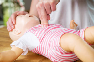
If you’re a bystander with no medical training, then you may not know how to effectively determine when to start CPR. In a CPR training course, you’ll learn what to look for.

2021 ILCOR guidelines continue to emphasize the importance of beginning CPR as soon as possible. So, as a general rule, if you suspect someone needs CPR because they’re unresponsive or gasping for air, you should begin CPR. If the person you’re giving CPR stops you with words or body language, that’s generally your best sign that they don’t need it.

Although they could be choking, in that case, your knowledge of first aid for choking would come in very handy. You’ll find a free 100% online CPR, AED, and First Aid course here. That lead
us to the next point.

A Broader Understanding of First Aid

Medical emergencies can take many shapes:

  • Burns
  • Chemical exposure
  • Allergic reactions
  • Severe asthma
  • Choking
  • Bee stings
  • Neck injuries
  • Uncontrolled bleeding
  • Trauma

CPR is important, as chances are you’ll need to administer it to someone, probably a loved one, in your lifetime. But you’re much more likely to need to manage an event that requires first aid several times in your life. These events can be life-threatening, like choking or anaphylactic shock. Or they may simply be a matter of reducing pain until a medical professional can examine the person.

Either way, every high school student up through adulthood needs to know First Aid.

What’s the Purpose of CPR and First Aid Certification?

We’ve established that you can perform CPR without formal training, so you also don’t have to become CPR certified. You can take a course for free online. You might also find a free course offered at a local high school, medical facility, civic center, or workplace.

Isn’t it great to know you can choose whether or not to get certified! And considering that this is a personal choice, here are some reasons to take that extra step to get certified in CPR and first aid.

CPR Is a Valuable Work Skill

Many people choose to go ahead and take that test to get certified because a CPR certificate looks great on your resume. When all else is equal, employers like to hire people who they can verify have CPR and first aid training.
Like good people skills or computer skills, CPR is a work skill and a valued one.

This makes the workplace a safer place to work.

Getting certified also shows personal initiative and commitment to lifelong learning. This may influence promotion and pay rate discussions that could lead to career advancement.

Some of the top workplaces where CPR certification would be very enticing to your employer include:

  • Schools
  • Daycare
  • Senior Centers
  • Home health / Hospice
  • Dental offices
  • Home service providers (HVAC, landscaping)
  • Construction
  • Warehouse
  • Law enforcement
  • Emergency services
  • Retail
  • Grocery
  • Restaurant
  • Entertainment venues
  • Any customer-facing business

Family Health

Make it official within your family. You’re CPR certified. This not only has a physical impact on your family health but can also provide peace of mind to those in your family who may have life-threatening health conditions like:

  • Heart disease
  • Respiratory diseases
  • Food allergies

Your certificate can deliver great comfort to your aging parents or grandparents. With you around, they’re less likely to see their lives cut short by a cardiac event.

Proof of What You Know

No one can legitimately say you don’t know CPR. You have proof in the form of a certificate.

Let’s face it. There’s something to be said when someone else recognizes your accomplishments and can verify that you’ve completed CPR training and know what you’re doing.

Maintaining Licences

If you have a medical license such as EMT or Nursing, then you may need Continued Medical Education Credits (CME) to maintain your license. For CPR training to count toward CME, you would need to take the test and get the certificate.

Why Aren’t More People Getting CPR Training?

Given the importance of CPR training, you might be shocked to find that 54% of people in the US do not know CPR in any form, according to a Cleveland Clinic study. Far fewer have actually had CPR training or obtained CPR certification.

Only 17% know that hand-only CPR (without mouth-to-mouth) has been proven as regular CPR for adults

Many people don’t realize how important CPR training is until they’re in one of these life-threatening situations. During an emergency, it’s too late to get that training so you can confidently save a life.

Others can’t find the time or don’t want to spend a whole Saturday in a high school gym learning CPR.

So it’s important to get the word out. You can learn CPR online in a Joint Commission-compliant course that teaches you all of the ILCOR-Approved CPR and First Aid methods.

These courses allow you to go at your own pace, and taking a course is 100% free. Simply complete the course, and at the end, you can choose if you want to take the step of getting certified. It’s up to you.

Have you already taken such a course? Share your experience with us.

About Greta

Greta is a dedicated life saver and a distinguished expert in the field of medical content creation and editing. Her impressive array of certifications in ACLS, CPR, PALS, and BLS underscores her commitment to excellence in the medical field. With over four years of invaluable experience in medical education, Greta plays an indispensable role within the Advanced Medical Certification team, shaping the way healthcare professionals around the world acquire and apply vital knowledge.

Greta's profound expertise serves as the driving force behind the development and distribution of medical content that has significantly enhanced the capabilities of countless healthcare practitioners across the globe.

In addition to her medical qualifications, Greta holds a prestigious academic distinction in Marketing and Global Business from Vilnius University. Her academic journey has been enriched by immersive studies in Slovakia and Portugal during her time as an exchange student, providing her with a global perspective that complements her medical expertise.

Beyond her professional commitments, Greta possesses a genuine passion for global exploration, with a particular focus on immersing herself in diverse cultures and appreciating the intricacies of the natural world. While residing in Vilnius, Lithuania, she continues to make substantial contributions to the field of medical education, leaving an indelible mark on the sector.

Reach out to Greta at greta.kviklyte@advmedcert.com.

Do This During a Severe Asthma Attack

Do This During a Severe Asthma Attack

Photo of Greta

by Greta Kviklyte

Life Saver, AMC
Co-authored by Kim Murray, RN, M.S.

posted on Oct 8, 2021, at 7:24 am

Share

If you or someone you know suffers from asthma, you’re familiar with common symptoms like:

  • Inability to draw a full breath
  • Wheezing
  • Chest tightness
  • Coughing

But not everyone experiences asthma the same way, and sometimes an asthma attack can be severe, requiring first aid and treatment of the restricted airways blocking proper function of the respiratory system. Would you know the difference? Read on to explore this topic and make sure you know what to do if someone you love has a severe attack.

Who Is at Risk of a Severe Asthma Attack

First of all, doctors classify asthma as mild, moderate, or severe based on how bad its symptoms normally are and how frequently they occur. Some people have intermittent asthma, where they occasionally suffer an attack.
Others have persistent asthma. For these individuals, asthma is an ongoing challenge. They may have attacks several times a day and need full-time treatment and first aid.

Anyone Can Have a Severe Asthma Attack

Anyone with asthma, even the mild kind, can have a severe asthma attack, often caused by something in the environment that causes a sudden flare-up or exacerbation of the “normally-experienced” asthma symptoms.

By normal, we mean normal for that person.

This event is life-threatening, and the symptoms will continue to escalate unless action is taken on either the part of the patient or the loved one.
During a severe attack, the airways become inflamed and contract to the point that little to no air can get through. The body also begins to over-produce mucous, intended to clear the airways of he irritant, but it ends up further blocking them.

If this person is not removed from the stimuli contributing to severe asthma (perfume, pollen, campfire smoke, cat/dog dander, etc.) and/or has access to their rescue inhaler, asthma can exacerbate to the point of severe asthma.

According to CDC.gov, a virus, such as a coronavirus responsible for COVID-19, can also trigger severe asthma in someone with mild to moderate asthma.

And, of course, anyone who has asthma found out they had it after their first attack, so even someone who has never been diagnosed with asthma could potentially experience a severe attack the first time.

Factors That Increase Risk of a Severe Asthma Attack

You are at increased risk of having a severe event if you:

  • Were first diagnosed after age 40
  • Have gone to the ER during an attack within the last 10 days. Severe attacks often happen near each other.
  • Take a steroid for your asthma. Note: the steroid itself doesn’t increase the risk. It’s the fact that you need one—correlation, not causation.
  • Use your rescue inhaler more than twice a month.
  • Have a co-existing condition, especially those of the heart or respiratory system

Signs of a Severe Asthma Attack

Student helping her asmathic friend giving the inhaler during an asthma attack
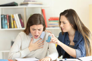
Doctors classify a severe attack as one that lands you in the emergency room or clinic. But before that happens, someone needs to recognize the person has a severe attack. Here’s what to look for:

Severe Wheezing

Severe wheezing is wheezing that a person can hear from across the room. It is the sound of air trying to force itself in and out of the airways. If this person has a rescue inhaler, they may be able to dilate their airways and stop the attack. But if they do not, this person may need first aid and treatment.

Uncontrollable Dry Coughing

The uncontrollable coughing is the result of a feeling of not being able to clear their throat. This cough usually sounds dry and tight, as opposed to wet coughs someone might have with a cold or flu that results in the person coughing something up.

Fast, Shallow Breathing

Because the person can’t get enough oxygen in one breath, they begin breathing rapidly, with each breath only pulling in a little air. Even with the rapid breathing, they’re unlikely to be getting enough oxygen to remain conscious, and that leads us to the next signs of severe asthma.

Chest Retraction

Normally, when you breathe, the chest retracts and releases with each breath. This requires muscle movement but also air pressure. The chest appears to cave in slightly during a severe asthma attack but doesn’t expand back out.

This is much easier to see in a baby or small child since their chests are smaller and still growing.

Panicked Feeling

This feeling of panic has two primary causes. First, if a person can’t breathe. That’s terrifying when the brain senses that it’s not getting enough oxygen, which provokes fear.

But the rapid breathing also activates the sympathetic nervous system that causes a fight or flight response. This causes the body to release adrenaline, which causes a panic feeling.

Anxiety and panic attacks can also trigger asthma in those who are susceptible to it, making this a vicious cycle in some.

Tight Chest

The person with asthma may cluck at their chest because it feels tight and painful.

Inability to Talk

A person having a severe asthma attack may try to speak, but sentences are cut short by quick breaths as they try to get out what they’re saying.

Blue Fingernails

The lips and fingernails turn blue first, so pay attention to those even if the face looks more or less normal.

Why does this happen? When the brain senses that the body isn’t getting enough oxygen, it will ration the little oxygen it gets to keep the brain, heart, and other vital organs working as long as possible.

That means non-essential extremities, fingers, toes, lips will go blue first.

Pale, Sweaty Face

The face may not turn blue immediately, but the person may start sweating, a natural fight or flight response from that earlier adrenaline release. You may also notice them getting paler.

These are signs of a severe asthma attack. Now, what should you do?

Creating a Severe Asthma Attack Action Plan

During an event, you certainly don’t have time to create an action plan. Instead, you’ll be thinking on your feet. But if you or a loved one even has mild asthma, creating an action plan for the two of you is a responsible step in the right direction.

  • Talk about signs of a severe asthma attack and when it’s time to call 911.
  • Talk to your loved one about known triggers. This is critical because if you need to move this person from the trigger, you need to know what you’re removing them from, if possible.
  • Make sure you know how to use an inhaler and where your loved one keeps it.
  • Learn the difference between respiratory distress and respiratory failure.
  • Learn CPR and First Aid for respiratory distress.

Note: People with chronic lung disease should have a rescue inhaler containing albuterol or another rescue drug. They may also have an attack prevention inhaler (montelukast sodium) that works by blocking the immune system’s production of certain inflammatory substances (leukotrienes), but this is not a rescue inhaler. It’s a preventative. Know the difference.

First Aid for Asthma: How to Use a Rescue Inhaler

woman-getting-an-inhalerIf possible, remove the person from the suspected irritant and find somewhere where they can get fresh air. Keep in mind, what you consider fresh air and what this person considers fresh air may be two different things. If this person is allergic to pollen, then taking them outside on a warm spring day may not be the best idea

  • Shake the inhaler 10 to 15 times. Note: If you know the person regularly uses this inhaler, then you only need to shake it a couple of times to prime it, according to medlineplus.gov.
  • Attach the spacer if available. Spacers deliver a slower, more continual dose which can be more effective than straight from the inhaler, especially if the person is struggling to take instructions.
  • Place spacer or inhaler spout in the mouth. It’s not like the movies where people use inhalers by holding them up to their lips. The part that releases the medicine is in the mouth, and the mouth is closed.
  • Press down on the inhaler while the person inhales deeply
  • If possible, they should hold the medicine for 10 seconds. If they’re already very short of breath, this may not be possible.
  • Repeat if symptoms continue
  • Stay with the person until medical assistance arrives
  • If this person becomes unresponsive, start CPR protocol.

CPR for a Victim of Asthma Attack

You only perform CPR on someone having an asthma attack which has stopped breathing and is non-responsive. You will follow standard CPR procedure as outlined in an ILCOR-aligned CPR training course. This should include rescue breaths.

Hands’ only CPR is normally acceptable if you prefer not to do mouth-to-mouth or don’t feel you can while doing compressions. Bystander hands-only CPR has been shown to be just as effective as CPR that includes rescue breathing, according to a review of three randomized trials published at nih.gov.

However, in the case of an asthma attack, the obstruction in the respiratory system has caused a lack of oxygen, so rescue breaths are essential if at all possible. One rescuer can give breaths and perform compressions with CPR training.

How Paramedics Handle a Severe Asthma Attack In the Field

According to JAMA, it takes seven to 14 minutes for an ambulance to arrive after you call 911. So, if your loved one is unresponsive, you should not wait to begin CPR. If they are still alert, you should begin first aid.

These could be the difference between life and death. Once emergency responders arrive, they will take the following actions.

Assessment

The EMT or paramedic will assess the situation.

This includes the ABC’s –airway, breathing, and circulation. Since they’ve been informed that this is an asthma attack, they will use an O2 Saturation monitor to get the victim’s oxygen levels. Carbon dioxide build-up is also a concern during an asthma attack, so they’ll attach another monitor to determine how much CO2 they’re breathing out (if they’re breathing).

If they’re still breathing, they’ll listen and watch for several of those signs of an asthma attack discussed in the above section. Is the person turning blue? Are they wheezing loudly? Have they tried their rescue inhaler?

If the person needs CPR when they arrive, they’ll stabilize the patient before proceeding.

This may seem like a lot of assessment steps, but emergency personnel can do all of this within seconds of arriving.

Provide Oxygen

Next, they’ll provide oxygen through a simple nasal cannula or facemask, which may work better if the victim is still struggling significantly.

Start the Nebulizer Treatment

A nebulizer is like a more powerful (and much larger) rescue inhaler. It may contain the same medicine but at a much higher dose and over a longer period of time. It delivers a steady flow of bronchodilation medication to stop the constricting in the respiratory system.

Albuterol is the main nebulizer medicine, but EMT may also add ipratropium to the machine. These two together are more effective than albuterol alone.

The victim stays on the nebulizer until they are no longer wheezing. A nebulizer can increase the person’s heart rate, which may already be fast because of the ordeal, so this is something they’ll monitor, although a racing heart will not be their primary concern.

Injections and IVs

In most cases, a little time on the nebulizer treatment resolves the issues. But emergency personnel must be ready for the possibility that the patient will destabilize. If they assess that there is a high risk of this, they may start a hydration IV and administer a steroid, such as methylprednisolone, which has been shown to reduce the need for admission due to severe asthma attacks.

They can also give steroids and other medications IM (muscle injection), if needed.

Airway Management

If the patient doesn’t respond to the above first aid and treatment, the next step is ventilation by way of a CPAP or BIPAP in the field. They will intubate (insert a tube) only if CPAP isn’t able to deliver air into the respiratory system.

Seeing the Doctor

The ambulance will typically take this person to the emergency room, where they will see a doctor. The doctor will examine the patient, make sure they understand how to manage their disease, and discuss prevention and treatment options. These may include being under observation or admission for a day or two.

First Aid for a Severe Asthma Attack

An attack is stressful for both the person having it and their loved one who helps them through it. If you have a friend or loved one at risk of a severe attack, then it’s critical that you learn CPR and first aid for asthma. Your actions could be the difference between life and death during these events. Share your experience of learning CPR and first aid with us.

About Greta

Greta is a dedicated life saver and a distinguished expert in the field of medical content creation and editing. Her impressive array of certifications in ACLS, CPR, PALS, and BLS underscores her commitment to excellence in the medical field. With over four years of invaluable experience in medical education, Greta plays an indispensable role within the Advanced Medical Certification team, shaping the way healthcare professionals around the world acquire and apply vital knowledge.

Greta's profound expertise serves as the driving force behind the development and distribution of medical content that has significantly enhanced the capabilities of countless healthcare practitioners across the globe.

In addition to her medical qualifications, Greta holds a prestigious academic distinction in Marketing and Global Business from Vilnius University. Her academic journey has been enriched by immersive studies in Slovakia and Portugal during her time as an exchange student, providing her with a global perspective that complements her medical expertise.

Beyond her professional commitments, Greta possesses a genuine passion for global exploration, with a particular focus on immersing herself in diverse cultures and appreciating the intricacies of the natural world. While residing in Vilnius, Lithuania, she continues to make substantial contributions to the field of medical education, leaving an indelible mark on the sector.

Reach out to Greta at greta.kviklyte@advmedcert.com.

How to Become a Trauma Nurse and Why You Should

How to Become a Trauma Nurse and Why You Should

Photo of Greta

by Greta Kviklyte

Life Saver, AMC
Co-authored by Kim Murray, RN, M.S.

posted on Sep 15, 2021, at 4:44 am

Share

A trauma nurse works in emergency wards and elsewhere, handling serious medical situations, some of which may be life and death. You’re trained to handle various minor and more severe trauma-related medical issues alongside doctors and other medical personnel.

You may provide care for a patient who experiences trauma from serious events like:

  • Accident
  • Gunshot
  • Domestic violence
  • Worksite mishap
  • Suicide attempt

According to the Centers for Disease Control and Prevention (CDC.gov), 35 million such patients come into the emergency room yearly. 43.5% of these trauma patients are seen within 15 minutes, and 12.5% will be admitted for ongoing treatment.

Trauma nurses help emergency departments and trauma units meet this demand.

What Does a Trauma Nurse Do?

healthcare professional attending a patientBecause of their injuries, these patients may experience strokes, heart attacks, internal bleeding, severe burns, and other conditions. Your training helps you identify and respond to these within your nursing capacity.

Trauma Nursing Care

To care for trauma patients, you’ll need advanced medical training like Basic Life Support BLS Certification and Advanced Cardiac Life Support ACLS Certification. These types of life-saving education help you work effectively with a medical team during emergencies to stabilize the patient and save their life.

Even after becoming a trauma nurse, you’ll be responsible for keeping your medical education current with continuing medical education credit (CME).

For example, with ACLS certification, as ACLS algorithms change, you and the other medical professionals on your team will all stay on the same page because you’re all up-to-date with ACLS training.

Studies show adherence to ACLS protocols improves patient outcomes.

Patient Education

You’re also uniquely qualified to provide the appropriate level of medical education to trauma patients and their families. You may need to explain more complex medical information in a way the non-medical person can understand.

You may teach family members how to cleanse, change bandages, and provide first aid for their loved one once they go home.

Compassionate Care

Trauma isn’t just about the physical damage. You also provide support and comfort for patients and their families while they’re dealing with what is undoubtedly a very emotional situation.

Such work requires intelligence, adaptability, compassion, and dedication. Do you have what it takes? Read on to learn more about this rewarding nursing specialty.

What is the Career Outlook for Trauma Nurses?

The Bureau of Labor Statistics (BLS.gov) says nursing employment is expected to grow by 9 percent by 2030. Currently, there are shortages of new nurses, especially among the Baby Boomers, many of whom have recently entered retirement.

The Baby Boomers are currently the second-largest living generation, next to Millennials.

As they move into retirement, we’re not only losing nurses due to retirement. We also have many people getting to the age where many people start to experience health decline or are at increased risk of serious injury while out there living the good life as an active senior.

In the midst of the anticipated nursing shortage, we’ve now experienced a pandemic, furthering the necessity for specialized nurses, including trauma nurses.

It’s critical that we have nurses to nurse individuals back to health.

What’s the Difference Between a Trauma Nurse and an Emergency Nurse?

If you’re an ER nurse, you’ll undoubtedly manage trauma patients. They go into the emergency department, where you triage them for the appropriate level of care.

Alternatively, a trauma nurse works in a department reserved for trauma patients. These patients may have come in through the ER. But now, they need ongoing trauma care.

This is what a trauma nurse working with a trauma team provides.

A trauma nurse may also work in the critical care unit of the emergency department. You may work with trauma patients as they come in, while other ER nurses work with patients experiencing non-traumatic illnesses.

Trauma nurses and emergency nurses are often drawn to the hustle and high-pressure environment of the ER and trauma unit, as it can be very stimulating and rewarding for a person who can manage stress effectively.

How Do I Become a Trauma Nurse?

heathcare-professionals-attending-a-patientTrauma nursing is a rapidly developing area of healthcare. It continues a trend toward specialization.

At one time, we just had nurses who were expected to do everything. Now, we have cardiac nurses, trauma nurses, pediatric nurses, and more who have a broad education but are also specifically trained to work in a certain unit.

But there are even niches within trauma nursing, like burn unit trauma nurses or the trauma nurses who work along with a trauma surgeon during a life-and-death surgery to repair an injury.

You don’t have to specialize as a nurse. But specialization may make more money because you have very in-demand and specialized skills.

When you’re hired in a trauma unit, leadership knows that you’re already trained and can fit right in with little additional training. That’s valuable to employers.

Steps to Becoming a Trauma Nurse

Step 1: Complete an Entry Level Nursing Program. You can become a trauma nurse after completing a two-year, three-year, or four-year program that results in either a nursing diploma or degree. Both are accepted.

Usually, you’ll get a degree as a registered nurse (RN) or Master of Science in Nursing (MSN).

Step 2: Take and Pass Your RN Exam. This is through NCLEX (National Council Licensure Examination – NCSBN.org) if you’re in the US or Canada. You must have an unrestricted RN license before you can apply to become a trauma nurse.

Step 3: Start Doing a Nursing Rotation. Try out different types of nursing environments to see which one speaks to you. You might rotate through pediatrics, cardiac, ER, etc., before settling on this career path.

Step 4: Work in a Trauma Unit. Once you’ve decided trauma nursing is for you, stay in a trauma unit working as a nurse. You will need two years at Trauma Nursing with around 1000 hours each year.

This may include working in a trauma unit, critical care unit, life flights, trauma surgical rehabilitation, or emergency, even though you’re not technically a trauma nurse yet.

Any of these will give you a good taste of what it’s like to be a trauma nurse. Here, you’ll support the trauma nurses and begin learning and doing what they do.

Step 5: Take Trauma-Related Classes. Step 5 runs concurrently with step 4. You’ll need to complete 30 hours of coursework. That’s roughly two years going part-time.

Step 6: Become a Certified Trauma Nurse. Apply for your certificate to be officially recognized as a trauma nurse. For example, you may become a Trauma Certified Registered Nurse (TCRN) certified by BCEN.org

Step 7: Take on More Responsibilities. As a Certified Trauma Nurse, you can continue to work in this capacity. But your role may expand. As you pick up greater responsibility, you will likely all see your pay go up. Certified trauma nurses may become:

  • Public educators about trauma and injury prevention
  • A nursing trainer within a hospital or unit
  • A nursing manager
  • A quality assurance auditor
  • An advocate for better training, funding, performance standards, etc. in Trauma Units
  • An advisor for a private company

Step 8: Keep your training up-to-date. As a trauma nurse, you’ll continually learn on the job. Technology, medicine, and the best practices built around them change. But you’re also required to get a certain number of continuing medical education credits (CME) during each license renewal period.

These help you stay current on industry-approved standards and procedures. Fortunately, this part is easy.

For example, you can get and update Advanced Cardiac Life Support ACLS Certification and Basic Life Support BLS Certification online to fulfill part of the CME requirement.

Step 9: Get an Advanced Certification. If you want to further advance your career at this point, there are many ways to do it. Becoming a trauma nurse practitioner is one way.

In addition to becoming certified in trauma nursing, you could become certified as a nurse practitioner and become a certified trauma nurse practitioner.

As a trauma nurse practitioner, you would take on a greater role in diagnosing, prescribing, and developing treatment plans. These are normally beyond the scope of practice in nursing. But not if you’re a nurse practitioner. If, as a trauma nurse, you want more responsibilities like these, NP could be a good career path.

Tips for a Successful Career

Here are some tips that can help you become the best trauma nurse you can be.

1. Find a Trauma Nurse Career Mentor

A mentor can provide thoughtful guidance in an often emotionally charged trauma environment. They can guide you on how to choose a professional career.

A good mentor knows the ups and downs of trauma nursing and can share their experience. Mentors are often the only people that will give you honest feedback about your performances before you develop bad professional habits that might fly under the radar of your manager.

To start, seek out a professional who has been a trauma nurse for a while and has achieved many things you want to achieve in your career.

2. Be Active in Trauma and Emergency Nursing Associations

Many employers will recommend this. But even if they don’t, take the initiative. You’ll learn so much and make connections.

Some groups to consider include:

  • The Society of Critical Care Medicine (SCCM.org)
  • Emergency Nurses Association (ENA.org)
  • American Association of Critical-Care Nurses (AACN.org)

3. Continue Your Professional Nursing Education

The only way to meet the continuously changing need for trauma care is through continuous learning.

Take advantage of a range of educational options as they become available to you through your employer.

As a trauma nurse, you may find that a varied work schedule makes it hard to attend scheduled classes, so look for flexible online options like online Advanced Cardiac Life Support ACLS certification and Basic Life Support BLS certification courses.

Stay up-to-date with ACLS algorithms to perform at your best for your patients.

4. Be Assertive and Never Be Afraid to Speak Up

You’ll be in some tense situations, and emotions may run high. Some people manage stress better than others. People who don’t manage stress well sometimes turn to bully behavior to get what they want.

Bullying is never okay. And often, people who tend to bully will do so until they realize you’re not going to take it.

Set clear boundaries, especially with trauma surgeons and your nurse superiors. Clearly and professionally communicate how they can effectively communicate with you to get things done.

If you’re ever in a situation with an aggressive patient or family member where you feel unsafe, remove yourself from that situation and get support.

5. Get Training in Leadership and Management Skills

Being a trauma nurse is a great pathway to leadership and management because you’re showing you can take the heat. You’re not afraid of uncertainty.

You can manage your stress levels and perform your duties professionally.

So, even if you’re just starting and leadership is way down the road for you, start working on skills now.

By learning these skills now, you can practice and perfect them long before you’re up for promotion.

6. Learn How to Communicate with Trauma Surgeons

Doctors often have their ways of communicating. Nurses need to pay attention to their communication style and try to adapt to it.

Don’t be afraid to speak up if you identify an issue. Nurses spend more time with patients. You’re often in a better position to spot something that’s not right that the doctor could miss. But it’s important to know how to communicate this observation professionally for the patient’s sake.

7. Keep It All in Perspective

Trauma nursing is fast-paced and rewarding for someone who can manage stress effectively. Find ways to maintain your work-life balance, so you can come to work, love what you do, and then leave it at work, so you can enjoy your life.

How have you become a trauma nurse? Share with us on social media.

About Greta

Greta is a dedicated life saver and a distinguished expert in the field of medical content creation and editing. Her impressive array of certifications in ACLS, CPR, PALS, and BLS underscores her commitment to excellence in the medical field. With over four years of invaluable experience in medical education, Greta plays an indispensable role within the Advanced Medical Certification team, shaping the way healthcare professionals around the world acquire and apply vital knowledge.

Greta's profound expertise serves as the driving force behind the development and distribution of medical content that has significantly enhanced the capabilities of countless healthcare practitioners across the globe.

In addition to her medical qualifications, Greta holds a prestigious academic distinction in Marketing and Global Business from Vilnius University. Her academic journey has been enriched by immersive studies in Slovakia and Portugal during her time as an exchange student, providing her with a global perspective that complements her medical expertise.

Beyond her professional commitments, Greta possesses a genuine passion for global exploration, with a particular focus on immersing herself in diverse cultures and appreciating the intricacies of the natural world. While residing in Vilnius, Lithuania, she continues to make substantial contributions to the field of medical education, leaving an indelible mark on the sector.

Reach out to Greta at greta.kviklyte@advmedcert.com.

Cardioversion vs Defibrillation: What’s the Difference?

Cardioversion vs Defibrillation: What's the Difference?

Photo of Greta

by Greta Kviklyte

Life Saver, AMC
Co-authored by Kim Murray, RN, M.S.

posted on Aug 26, 2021, at 8:30 am

Share

Cardioversion vs defibrillation are two potentially life-saving methods that are critical for all rescuers to understand. If you are facing the need to provide synchronized cardioversion vs defibrillation or how to treat an irregular heart rate, read on.

Each year, an estimated 300,000 cardiac arrests happen in U.S. hospitals, of which some 81% are non shockable rhythms (asystole or pulseless electrical activity). In 50-60% of cases, cardiac arrest happened because of primary heart failure, followed by primary respiratory failure at somewhere over 15%. This is according to a JAMA study published at NIH.gov.

The only proven rhythm method to improve the survival rate to discharge is timely defibrillation after a cardiac arrest, secondary to ventricular tachyarrhythmia.

However, for certain types of cardiac arrhythmias, the most effective treatment is synchronized cardioversion. So it’s important to know the difference between cardioversion and defibrillation, when they’re used, and when one or the other may be dangerous and even cause cardiac arrest.

Synchronized Cardioversion Vs. Defibrillation: Overview

The goal of both defibrillation and cardioversion is to transfer electrical energy to the heart. This stuns the heart instantly in hopes of generating a normal sinus rhythm from the heart’s natural pacemaker, which the heart can then maintain on its own.

doctor-nurse-holding-defibrillator

While both use the same general action and can use the same equipment, the method is different, as well as when you use them, how you use them, and why you’re shocking the heart.

Primarily, you use defibrillation during an immediate life-threatening situation. You have a cardiac arrest, and if you don’t get this heart restarted, the victim will officially transition.

Conversely, doctors use cardioversion to convert heart rhythm when a patient has an unstable heart but is not immediately in danger of dying. Cardioversion stabilizes the heart rhythm. It is, therefore, usually an elective procedure. With guidance from their doctor, a patient chooses cardioversion to correct their heart rhythm. But it can also be urgent if the patient is experiencing tachycardia or a feeling of breathlessness.

Second, doctors synchronize cardioversion to achieve a specific rhythm outcome. You don’t time defibrillation. You just need a shockable rhythm.

So, as soon as there is a shockable rhythm, an automatic defibrillator tells you to “hands off” or “clear”. Then a shock occurs. But during cardioversion, the machine waits a couple of seconds to sync up with the rhythm to shock at a precise time in that rhythm.

Third, a defibrillator delivers a higher energy dose vs. cardioversion.

Ultimately, cardioversion performed when needed could prevent a subsequent cardiac arrest later that day, week, or year. That’s because it transforms an irregular rhythm into a stable one. But once a cardiac arrest occurs, defibrillation is your only option–that is, if you have a shockable rhythm.

Below you’ll find further information about these procedures to further distinguish between them.

Cardioversion vs Defibrillation: A Chart for Easy Comparison

Use the following chart to help you see the differences between cardioversion and defibrillation. Then, read on about the process of providing care.

Defibrillation vs Cardioversion

Category Defibrillation Cardioversion
Patient Condition The patient is in cardiac arrest or has no heartbeat The patient is functional but has an irregular heartbeat
Energy Levels Between 150 and 360 joules Between 50 and 300 joules
Synchronization Is done as necessary Set to a specific point according to an EKG
Sedation Requirement Midazolam or Diazepam Midazolam or Diazepam
Typical Arrhythmias Treated Life-threatening arrhythmia Used to convert an arrhythmia back to sinus rhythm, such as with atrial fibrillation

What Is Defibrillation?

Defibrillation is a shock-based treatment for life-threatening arrhythmias when a patient does not have a pulse. These could include ventricular fibrillation (VF) or pulseless ventricular tachycardia (VT). It’s done when someone is in cardiac arrest.

Someone can perform it in a hospital, an ambulance, or an out-of-hospital setting. While medical professionals perform defibrillation in the hospital, the average person with CPR training can also perform defibrillation if they have access to an automatic external defibrillator (AED). You can buy one for your home or office.

A Brief History of the Defibrillator

Two physiologists (Prévost and Batelli) discovered the power of fibrillation around the end of the 18th Century by running dog experiments using shock. They found that when they applied small shocks to the dog’s heart, they could put the heart into V-fib and then shock it again with a larger shock to reverse the change.

But it would be another 14 years before a cardiothoracic surgeon did defibrillation on a 14-year-old boy on whom he was performing cardiothoracic surgery for a congenital heart disorder. He placed electrodes on the open heart for the procedure.

The technology remained mostly unchanged for another 150+ years until the 1950s, when Russian researchers developed a defibrillator that they could use on a closed chest. This would later become the modern-day monophasic defibrillator. In the 1980s, researchers discovered a biphasic waveform, which would replace monophasic shocks in some instances.

Defibrillation and Cardioversion share this history since they both use a defibrillator to work.

Monophasic Vs. Biphasic Shock

A monophasic shock only travels in one direction from one paddle to another, while a biphasic shock travels from one paddle to the other and then back several times. Biphasic shock results in fewer burns and has a higher first shock success rate (90% to 60%), although studies comparing how use may impact discharge success haven’t been thoroughly studied.

Types of Defibrillators

There are four primary types of defibrillators, which are used in different settings:

  • Automated External Defibrillators (AEDs) are often found in CPR kits for the average person with CPR training.
  • Semi-automated AEDs can be overridden and are therefore only used by a trained professional like a paramedic.
  • Standard defibrillators with monitors require professional skills to operate. You’ll find them in hospitals mostly.
  • Implanted defibrillators can shock the heart back into a rhythm during a cardiac arrest without assistance from a bystander.

While paddles were once very common, they’ve now been overtaken by the use of adhesive patches, which are generally easier to use and perform better because they adhere to the chest without the need for gel application.

Risks of Defibrillation

Defibrillation risks are minimal when the machine is well-maintained. Defibrillators measure the patient’s heart rhythm and, if you have an automatic machine, they provide clear instructions based upon that rhythm. All you have to do is follow his instructions even if you have no medical training.

Fragile patients, like the elderly, may experience broken ribs or the aggravation of an existing injury. But defibrillation saves a life, so those are generally considered reasonable casualties of the procedure.

Shocking yourself is also possible, so when the machine says “hands off”, please make sure no one’s touching the victim.

Defibrillator burns have been reported but are rare with current equipment and a sign of a poorly-maintained machine that probably needs to be replaced. The batteries and pads of an automatic defibrillator typically last two to five years, depending on the frequency of use. So they need to be replaced to maintain proper function. An implanted defibrillator lasts up to 10 years, according to Johns Hopkins at HopkinsMedicine.org.

These same risks will apply to cardioversion plus a few more.

Energy Levels for Defibrillation

  • Monophasic – CPR algorithm recommends single shocks started at and repeated at 360 Joules (J).
  • Biphasic – CPR algorithm recommends shocks initially of 150-200 J. Then use subsequent shocks incrementally 150, 200, 300, 360 J.

A clinical trial on biphasic defibrillation showed that increasing the energy level of shocks was more likely to result in a conversion and stop ventricular fibrillation (VF) when compared with repeated shocks at 150 J. Often, the first shock did nothing, but the subsequent higher J shock did.

What Is Cardioversion?

man-lying-with-aed-padsSynchronized cardioversion is a process to achieve a sinus rhythm in a patient who has arrhythmia. It requires the delivery of a low energy shock at a specific time. Doctors achieve this timing by viewing a defibrillator monitor or using a machine that syncs to the rhythm automatically. In both cases, you’ll only find this equipment in a hospital or specialized clinic.

They sync the shock to a specific point in the QRS complex (the main spike visible on that monitor with EKG / ECG).

Through this method, atrial fibrillation can be converted to a normal sinus rhythm while preventing the prompting of ventricular fibrillation (VF). This happens because you’ve synchronized the electrical shock to the R wave. At the same time, you avoided that susceptible T-wave and averted ventricular fibrillation.

Uses & Indications

Patients who need cardioversion experience the following symptoms:

  • Breathing struggles from pulmonary edema (fluid in the lungs)
  • Hypotension (low blood pressure)
  • Chest pain
  • Syncope (loss of consciousness from low blood pressure)

When a patient has symptoms like these, their doctor will need to run tests to confirm the cause of the symptoms.

Electrical cardioversion may be indicated if those tests show:

  • Unstable pulse
  • Failed chemical cardioversion with chest pain or unlikelihood chemical cardioversion would be successful.
  • Decompensated rapid AF with rapid ventricular response. An example of this would be a hypotensive patient who isn’t responding to other medical therapies.
  • Ventricular tachycardias (VT) with pulse
  • Supraventricular tachycardias, which includes AF without decompensation but only if not in an acutely urgent situation.

The technique is used less frequently in situations such as atrial fibrillation to revert the heart rhythm to a sinus rhythm.

In a patient with atrial fibrillation, you use cardioversion to control the rhythm. However, cardioversion will not always be successful. Studies have shown as many as 50% of patients’ hearts will return atrial fibrillation within 12 months. However, a doctor could do another cardioversion at that time, if needed.

Risks of Cardioversion

In addition to the general risks of defibrillation, you should stay aware of these potential complications.

Having cardioversion may put patients at increased risk of thromboembolic disease (TED) soon after the procedure. As a precaution, doctors prescribe anti-coagulation at least three weeks before and four weeks after the procedure.

Doctors may also use a transesophageal echocardiogram during the procedure to spot thrombus, although some patients may still develop TED.

How to Cardiovert

Group of people training on aedCardioversion is performed in a hospital with a doctor, an anesthesiologist, and cardiac nursing professionals present. The anesthesiologist will plan and supervise sedation of the patient using general anesthesia. Once sedated, the team performs the following steps:

  1. Attach the self-adhering electrodes
  2. Pick an energy level to start. Note: a higher starting energy may require fewer shocks. Levels are based on the starting type of rhythm. For Broad complex tachycardia and AF, you’d do monophasic starting at 200 or biphasic at 120-150 J. For atrial flutter and narrow complex tachycardia, the recommendation is starting at 100 for monophasic and 70-120 J for biphasic
  3. Achieve a visible trace on the monitor
  4. Hit the sync button. You’ll see a blip or dot marking the QRS complex. This will allow the shock to sync automatically so it shocks with correct timing that might not be achieved manually. Note: sync may not work in tachycardia where the QRS complex is variable.
  5. Charge
  6. Call “Clear” and look to see that the bed is clear.
  7. Shock. Expect a 1-2 second delay since the machine is synchronizing the shock.
  8. View the rhythm to see if you have a sinus rhythm. If yes, stop. If not, adjust the energy level up incrementally and repeat steps 5-8.
  9. Look for burns that may need treatment
  10. Get a 12-lead ECG for a more thorough reading

Special Considerations on Infants and Children

Pediatric Cardioversion and Defibrillation require attention to detail and some basic math skills. The recommendation is to start at 1J for every kilogram of weight.

You should never use an automatic or semi-automatic defibrillator on a child younger than one year. Only use a manual one with a monitor, which usually means the child is in the hospital.

Whether performing on an infant or child, note that manual defibrillators only go as low as 4J/Kg, so use specialized pediatric pads for both manual and automatic external defibrillators. This reduces the energy delivered when used in combination with the pediatric setting. You can view further details related to pediatric treatment in Pediatric Cardiac Arrest Algorithm.

Children eight and older can receive cardioversion and defibrillation at adult levels, according to the guidelines outlined in your Advanced Cardiac Life Support Training. You can now obtain this training 100% online.

Pediatric defibrillation is complex in that the recommended shock level is much lower, beginning with a dose of 2 J/kg and use of 4 J/kg for subsequent attempts. Because ventricular fibrillation occurs in 8 to 20% of pediatric cardiac arrests, it is critical to consider the best treatment possible when administering this shock. Data indicates that, with every 1 minute of delay in defibrillation, the survival rate of the child falls by 7 to 10%.

Cardioversion Vs. Defibrillation

Cardioversion and defibrillation both rely on the power of shock to achieve the desired heart rhythm. But when they’re needed and how doctors use the defibrillation equipment are very different. Exploring the differences between procedures with so many similarities can help you better understand how the heart works and how doctors can apply different strategies to help the heart perform at its best.

Chemical vs Electrical Cardioversion

In the comparison of cardioversion vs defibrillation, consider the mechanisms used. Studies indicate that chemical vs electrical cardioversion can be a critical factor for doctors and rescuers to consider, and no single consensus exists on which method is the preferred option.

In short, a chemical cardioversion occurs when an abnormal heartbeat is cardioverted to a normal rhythm using medications. These medications work to get the heart back into normal rhythm. By comparison, electrical cardioversions use electrical shock to adjust the rhythm of the heart.

Both methods may be effective, and electrical cardioversion may be faster at converting a person. For those who cannot be shocked, the use of medication may be necessary.

Note that in cardioversion vs defibrillation, only cardioversion allows for a true chemical treatment. Defibrillation only occurs with the application of electrical energy, though both methods may be used during this process.

Synchronized Cardioversion vs Defibrillation

emt holding an aedYou may hear a significant amount about synchronized cardioversion vs defibrillation or defibrillation vs synchronized cardioversion. Let’s break down some of the core elements of this comparison of cardioversion vs defibrillation:

  • Synchronized cardioversion: This process occurs in a specific part of the heart’s beat cycle. More precisely, the first step is to recognize the electrical impulses that control the heartbeat and then measure them accurately. This allows for a jolt to be administered at a very specific time within the heart’s beating cycle, which could improve cardioversion outcomes. These cardioversions are timed.
  • Unsynchronized cardioversion: These cardioversions require the use of more energy because they are untimed and, therefore, can deliver a shock at any time during the cardiac cycle. This type of cardioversion is much more in line with a defibrillator.

In many cases, an unsynchronized cardioversion is the same application and treatment process as a defibrillation. It occurs at any time after the machine charges and is ready to fire, even without any monitoring of the heart’s beat.

How to Cardiovert in the Field

Cardioversions happen in a hospital setting. However, if there is an AED available, rescuers can use it with proper education. Rescuers can learn how to cardiovert using an AED after taking a licensing exam. From the objective of the patient, the process will include:

  • Being under twilight sedation
  • A cardioverter machine will deliver a quick jolt of energy to the patient. This energy, delivered to the heart through the placement of cardioversion pads, changes the beat.
  • It interrupts the abnormal beat or electrical rhythm of the heart and then restores normal heart rhythm, which is more natural and what the heart wants to beat.

In some situations, it may take several shocks to restore the normal function of the heart. The procedure only takes a few minutes. If you are a rescuer and wish to learn how to administer a cardioversion or defibrillation in an emergency, complete the CPR, AED & First Aid Certification Course we offer.

What is atrial fibrillation?

Atrial fibrillation is often a component of cardioversion vs defibrillation conversations. It indicates the heart’s quivering action or irregular heartbeat in the atrium or the upper chamber of the heart. Often called Afib, this condition is not usually life-threatening, though it can prolong complications to the heart and lead to complications over time.

Which is better, unsynchronized cardioversion vs defibrillation?

An unsynchronized cardioversion is the same as defibrillation. These types of cardioversion do not have a timed or planned moment of transferring the energy to the other patient. Rather, as soon as the device is charged up, it is applied without paying attention to the actual cardiac cycle.

What are synchronized cardioversion joules?

Synchronized electrical cardioversions typically utilize a recommended energy level of 50 to 300 joules, depending on the person’s age and overall condition. A narrow regular level indicates joules of 50 to 100 joules. If a wide regular heartbeat occurs, 100 joules are used. With a narrow irregular rate, 120 to 200 joules are used. What is the difference between synchronized and unsynchronized cardioversion? Synchronization indicates that the jolt of energy is applied at a very specific point in the cardiac cycle or the process of each heartbeat. This can help to change the heart rate back to normal rhythm without the use of as much electrical shock as what occurs in untyrannized cardioversions.

Ready to Learn More?

Enroll in the ACLS certification course now. Doing so allows you to learn more about cardioversion vs defibrillation and teaches you how to take action during an emergency medical event.

About Greta

Greta is a dedicated life saver and a distinguished expert in the field of medical content creation and editing. Her impressive array of certifications in ACLS, CPR, PALS, and BLS underscores her commitment to excellence in the medical field. With over four years of invaluable experience in medical education, Greta plays an indispensable role within the Advanced Medical Certification team, shaping the way healthcare professionals around the world acquire and apply vital knowledge.

Greta's profound expertise serves as the driving force behind the development and distribution of medical content that has significantly enhanced the capabilities of countless healthcare practitioners across the globe.

In addition to her medical qualifications, Greta holds a prestigious academic distinction in Marketing and Global Business from Vilnius University. Her academic journey has been enriched by immersive studies in Slovakia and Portugal during her time as an exchange student, providing her with a global perspective that complements her medical expertise.

Beyond her professional commitments, Greta possesses a genuine passion for global exploration, with a particular focus on immersing herself in diverse cultures and appreciating the intricacies of the natural world. While residing in Vilnius, Lithuania, she continues to make substantial contributions to the field of medical education, leaving an indelible mark on the sector.

Reach out to Greta at greta.kviklyte@advmedcert.com.

PEA Arrest Uncovered: The Vital Steps to Save a Failing Heart

PEA Arrest Uncovered: The Vital Steps to Save a Failing Heart

Photo of Greta

by Greta Kviklyte

Life Saver, AMC
Co-authored by Kim Murray, RN, M.S.

posted on Aug 9, 2021, at 11:36 am

Share

What Is PEA Arrest? Pulseless electrical activity (PEA) is also known as electromechanical dissociation. It’s a clinical condition in which a patient experiences unresponsiveness in conjunction with a pulse that cannot be felt even when applying sufficient electrical discharge.
The electrical impulse is relevant but not sufficient to restart the heart because something else is going on in a PEA arrest.

How Common Is PEA Arrest?

PEA arrests are a surprisingly common occurrence in the hospital setting.

A study reported in NIH.gov found that 68% of tracked in-hospital deaths and 10% of all in-hospital deaths could be attributed to pulseless electrical activity.

A PEA arrest is the first documented rhythm in as many as 38% of adults experiencing hospital cardiac arrest (IHCA).

Certain medications such as beta-blockers and calcium channel blockers may alter ventricular contractility. This leads to an increased chance of PEA arrest happening, and it is less likely that treatment will be successful. PEA arrest is most common in women, and the chance of this happening increases for those over 70, particularly among women.

How It Happens: Understanding the Dying Process

old-woman-holding-her-chestIn order to understand how to save a life during a cardiac arrest, you need to take a look at the dying process. This begins with the loss of function of a vital organ, e.g., brain, heart, or lungs. If professionals cannot restore this organ, it causes other organ failures.

However, in the case of a PEA arrest, the vascular system has collapsed. Without the vascular system, that blood can’t get to other vital organs, so the brain and lungs stop. In reality, the vascular system should also be considered a vital organ. Now death begins. The heart continues to pump until it no longer has the oxygen it needs for cardiac functions. Loss of effective pulse happens next. This loss of pulse is the beginning of PEA arrest.

So a PEA arrest is not a primary cardiac arrest. It is a latter stage in the dying process that begins with the stopping of another vital organ: the brain, the lungs, or the vascular system for one reason or another.

Examples of PEA Arrest Causes

Hypoxia is one of the most common causes of PEA, leading to an estimated 1/2 of PEA events. With oxygen cut off, the heart no longer has the means with which to contract, even if the heart were otherwise fully capable of a contraction with electrical stimulation from an AED. Unless the oxygen is restored, the heart muscle will die, and death results. This patient probably needs intubation before shock will have an effect.

The same can occur with:

    • Decreased preload from hypovolemia (loss of blood impairing atrial contractions)…This is the second most common cause of PEA.
    • Increased afterload from something like vascular resistance
    • Poor contractility, usually caused by acidosis or a reduction in calcium concentration

Primary Causes of PEA

Pulseless electrical activity, the PEA medical abbreviation, can be caused by several factors. As noted by the Cleveland Clinic, the most common possible causes include:

  • Problem with the function of the heart
  • Blood loss or low blood pressure
  • Severe dehydration
  • Imbalance of electrolytes
  • Heart attacks
  • Low oxygen levels
  • Hypothermia (low body temperature
  • Trauma to the heart or the chest area
  • Air, blood, or fluid fills the internal chambers of the body, including the chest, applying pressure to the heart.
  • Pulmonary embolism
  • Toxic effects from prescription or recreational drugs

Note that pulseless electrical activity can occur from one or a combination of the above factors.

What Are The Reversible Causes of Cardiac Arrest (H and T)?

Experts break down the reversible causes of cardiac arrest into two primary categories, conveniently called H and T, or H’s & T’s. Any of the H and T may lead to a PEA.

The H’s are:

  • Hypovolemia – Loss of blood volume (bleeding out)
  • Hypoxia – Loss of Oxygen
  • H+ (acidosis) or Metabolic Acidosis, an increase in hydrogen
    concentration in the body leading to a low serum bicarbonate
  • Hypo/Hyperkalemia – Too little / too much potassium in the body
  • Hypoglycemia – Low blood sugar
  • Hypothermia – Low body temperature

The T’s are:

  • Tension pneumothorax – ongoing entry and trapping of air in the pleural area around the lungs
  • Tamponade (Cardiac) – Pressure caused by fluid or blood building up in the area outside the heart muscle in the heart sac
  • Toxins – These could include sedatives, opioids, pesticides, acid, anaphylactic shock-inducing allergens, sodium-potassium blockers, etc.
  • Thrombosis (pulmonary embolus) – A blockage, usually a blood clot that gets caught in the lungs
  • Thrombosis (myocardial infarction) – Usually a blood clot in the vessels of the heart
  • Trauma (physical) – A serious bodily injury, e.g., blunt force trauma or penetrating trauma

How Is PEA Treated?

If the PEA arrest is among the H and T, then it may be reversible. But you must treat the cause of the PEA arrest to reverse the state and obtain a shockable rhythm.

In a hospital setting, this may involve several Advanced Cardiac Life Support Techniques and procedures such as:

  • Needle decompression of a collapsed lung
  • Laryngeal tube / Intubation
  • Blood infusion
  • Body temperature correction
  • Surgery to remove the pulmonary embolus
  • Epinephrine

With that said, since hypoxia accounts for over 50% of PEA, epinephrine and ACLS airway management such as intubation are the emergency procedures stated in the ACLS Cardiac Arrest Algorithm. These measures are more about buying time so that the cause can be addressed. That may require surgery.

According to the algorithm, if you have a shockable rhythm, you shock before intubating the patient. But if they have a non-shockable rhythm (PEA/Asystole), you prioritize intubation without restoring oxygen, since the heart will stay unshockable until you do.

Treatment for PEA in ACLS

Following the PEA algorithm enables rescuers to take the necessary steps to provide PEA medical help in a critical moment. According to ACLS guidelines, the treatment requires:

  • High-quality CPR
  • Establishment of an IV access
  • Administration of epinephrine at a rate of 1 mg every 3 to 5 minutes
  • Work to identify and treat reversible causes

At the time of diagnosis, treatment should be specific management of that concern. This could include:

  • Correction of the body’s temperature
  • Pericardial drain for tamponade
  • Fluid infusion for dehydration
  • Decompression for pneumothorax
  • Administration of thrombolytics for PEA cardiac arrest

Treatment for PEA rhythm is always specific to the underlying concern. However, epinephrine should be given through an IV on an ongoing basis, every 3 to 5 minutes, when PEA cardiac arrest occurs. Follow this with a 20 ml flush and then elevate the arm for 20 seconds to improve perfusion.

What’s the Difference Between PEA and Asystole?

The two are related cardiac rhythms, since they are both potentially deadly and non-shockable rhythms, requiring intervention before you can shock.

An Asystole is a flat line ECG, so you may have subtle movement away from the baseline (a drifting flatline). But you cannot perceive the cardiac electrical activity. A PEA is also one of many waveforms with no detectable pulse on ECG. Per International Liaison Committee on Resuscitation (ILCOR), any pulseless waveform can be called PEA, except ventricular fibrillation (VF), Ventricular tachycardia (VT), and Asystole.

An Asystole usually occurs because of trauma or accidental shock (touching a live wire, lightning, etc.), which stuns the heart muscle, causing the asystole. This can lead to an imbalance in the electrolytes sodium and potassium on the inside and outside of heart cells. Without those electrolytes, the heart cannot pulse or pick up on electrical impulses.

Because of the nature of the disruption, the heart experiencing Asystole no longer has the means to move an electrical current generated by defibrillation through the heart. It is therefore unshockable.

IV fluids can help restore this balance and are therefore another part of the ACLS cardiac arrest algorithm.

What Does an Asystole Look Like on ECG?

On ECG,

  • The rhythm will have an almost flat line appearance
  • No rate
  • No P-Wave, so you cannot measure PR interval
  • No QRS complexes

Pulseless Electrical Activity1
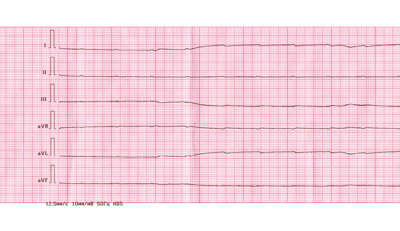
What Does a PEA Look Like on ECG?

  • There may be any rhythm, which includes a flatline
  • Any rate or none
  • Possible P wave or none
  • Possible PR or none
  • Possible QRS complex or none

What Are Shockable vs. Non-shockable Rhythms?

A shockable rhythm is one caused by abnormalities in the electrical conduction in the heart. These include:

  • Ventricular Tachycardia
  • Ventricular Fibrillation
  • Supraventricular Tachycardia

By addressing the cause of the PEA arrest, you can return the heart to one of these shockable rhythms. It’s critical that healthcare professionals stay vigilant when a PEA or Asystole converts back to a shockable rhythm. You can now use a defibrillator to shock and continue to follow your ACLS algorithm.

How Is PEA Diagnosed?

Pulseless Electrical Activity ECG Paper
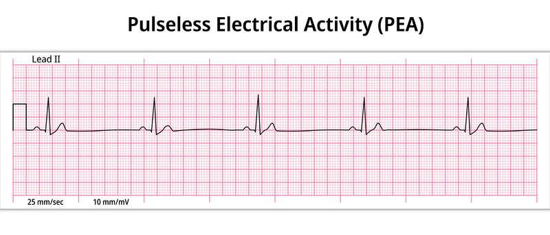
The first step in PEA arrest is action. The only way to know if a heart arrest is occurring as a result of a PEA is through the use of an electrocardiogram. Most of the time, electrocardiograms are not readily available outside of a medical facility, which makes it hard for rescuers to know that a PEA arrest is occurring.

However, knowing what PEA arrest is is not as important as the actions that a rescuer takes at that moment. Since the treatment for PEA is the same as it is for cardiac arrest, it is not as important to know what is occurring initially. When this can be assumed, it may be possible for providers to work to reverse the underlying cause of the condition.

What Is Pseudo-PEA?

The term Pseudo-PEA indicates that an organized electrical cardiac rhythm is present and there is cardiac muscle activity, however, there is no palpable pulse. This occurs due to profound shock. Typically, the PEA ECG or echo will show that there is muscle activity and a cardiac rhythm but without the proper functionality to sustain life.

What’s the Difference Between PEA Arrest and Pseudo-PEA?

Pseudo-PEA and PEA both have organized activities that you can see on the monitor. And in both cases, you can’t detect a pulse using manual palpitations.

The defining factor is that in PEA arrests, there really is no pulse. There is no cardiac output. There is cardiac activity, but for some reason that activity is fruitless.

However, in the case of a Pseudo-PEA, even though a manual test detects nothing, you can detect a pulse by other means: arterial line, POCUS pulse check, ETCO2, and oxygen saturation waveform.

Why does this matter? Because a Pseudo-PEA may be shockable because it’s probably one of the three shockable rhythms. But depending on the devices used to determine “pulselessness”, one might falsely determine a PEA arrest and not shock.

What Happens If You Shock PEA? Why not shock a PEA Arrest?

In a PEA arrest, similar to Asystole, the heart doesn’t have the means to use the shock you’re sending it because the primary cause has yet to be corrected.

Shocking a heart in PEA arrest is like kicking a comatose patient in the abdomen (which we do not recommend). The kicking may move them around and cause what seem to be signs of life. But it doesn’t wake them up. It’s more likely to cause damage than help the situation.
With that said, the occurrence of Pseudo-PEA is common, so before making the decision not to shock, check your equipment, placement of leads, etc. to confirm PEA.

Can You Survive PEA?

Yes, you or your patient can survive PEA if you eliminate the primary cause of the PEA arrest to return the heart to a shockable rhythm. Then resume actions according to the ACLS cardiac arrest algorithm.

What Is True PEA?

In comparison to pseudo-PEA cardiac arrest, true PEA is called electro-mechanical dissociation. It occurs when organized electrical cardiac rhythms are present. However, no cardiac muscle activity is present. This indicates that the muscle (heart) is not moving. PEA arrest does not involve any muscle function evidence on an ECG.

Which Medications Are Considered First for PEA?

Current ILCOR guidelines suggest that atropine is not to be used on PEA or Asystole. There is no evidence that it works, but there is also no evidence that it’s harmful.

Although research is ongoing in this area, ILCOR leans toward high-dose epinephrine. In limited studies, this has improved ROSC (Return of spontaneous circulation). With that said, there is some debate about how much epinephrine to administer, as some studies have shown no added benefits for administering higher than 1mg.

ILCOR has also suggested the use of a vasopressor, conveniently named vasopressin, which could replace the epinephrine or be used in conjunction with it, since Epinephrine loses additional effectiveness after 1mg. However, the two are more or less equal in effect, so for simplicity, you’ll only see epinephrine in the ACLS algorithm.

Some other medications to consider based on the cause of PEA and whether you can resume a shockable rhythm include:

  • Adenosine
  • Amiodarone
  • Atropine
  • Dopamine
  • Lidocaine (only if Amiodarone isn’t available)
  • Magnesium Sulfate
  • Procainamide
  • Sotalol

PALS PEA Arrest Management

running-doctors-and-nursesFor pediatric patients, you have a separate PALS cardiac arrest algorithm. The general flow for PALS PEA management is the same as that of an adult with the exception of PALS-specific CPR techniques and child-appropriate medication doses.

7 Tips for Managing a PEA

1. Recognize the Challenge

When a patient presents with a PEA arrest, your resuscitation team has a challenge ahead. This isn’t as straightforward as a shockable rhythm.
But follow your ACLS training for the scientifically best chance of success with a PEA arrest patient.

2. Know That Survival Rate Is Low

As medical providers, we always want to go in with a positive and practical mindset. But the truth is that, since PEA arrests are harder to manage, the survival rate is lower than if your patient came in with a shockable rhythm.

3. Think Of Your Team As the Holding Team

As a resuscitation team, you’re trying to buy your patient time for other medical treatments to work. The ACLS algorithm doesn’t solve the causes of PEA arrest. But it can lead to ROSC and stabilization, which gives a surgeon more time to address the issue and save the patient.

4. Know Your H and T

This mnemonic can help you remember an otherwise long list of causes of PEA that may be reversible.

5. Pay Attention to the Rhythm on ECG

Timing matters. When that rhythm becomes shockable, your team should be ready to shock all while continuing CPR and the other steps covered in the cardiac arrest algorithm.

6. Check Medical History When Available

You can often more quickly narrow the list of possible causes of PEA arrest if you know their medical history.
The patient may have had risk factors for hyperkalemia (electrolyte imbalance) or they may have recently left an addiction treatment facility.
Pulling this history may be a role for someone on your in-hospital code blue team.

7. Make Sure Team ACLS Training Is Up-to-Date

It’s critical that your whole code blue team works from the same playbook. Great teams think alike and follow a shared mental model (protocol).
Staying up-to-date with ACLS no longer requires spending a Saturday in a classroom. You can complete ACLS and PALS courses 100% online and get certified online too.

A PEA Arrest

A PEA arrest is a serious business, and your team will certainly have an uphill battle to ROSC. But through ACLS training, you can learn how to manage a PEA Arrest and improve the outcome for many patients.

Prognosis of PEA

PEA arrest often leads to a poor outcome. In one study of 150 patients, about 232% of those who had PEA arrests were resuscitated and survived until they could be admitted to the hospital. Additionally, of those who had a PEA arrest and were admitted to the hospital, only 11% survived until discharge. A high mortality rate exists in PEA cardiac arrest.

What is your experience with treating PEA arrest? Share with us on social media.

PEA Arrest FAQ

Is PEA a Shockable Rhythm?

PEA arrest is not considered a shockable rhythm. This means that defibrillation is not an appropriate or effective treatment option. However, rescuers should start CPR to restart cardiac activity. If a shockable arrhythmia, such as ventricular tachycardia, is present at that point, then the administration of a defibrillator can be effective.

What Is PEA Arrest?

PEA arrest occurs when there is some electrical activity within the heart, but it is too weak to provide enough contraction to support the heart’s ability to beat. Typically, the pulse is not possible to detect in these situations.

What is PEA?

PEA stands for pulseless electrical activity. It is a type of arrhythmia associated with the lack of effective electrical activity in the heart. It is a highly dangerous type of cardiac arrest requiring immediate medical care.

What is asystole vs PEA?

PEA occurs when there is some detectable electrical activity. However, with asystole, no electrical activity is present or detected.

Key takeaways:

  • PEA arrest occurs when a person’s heart is not functioning normally. In PEA arrest, the heart has some detectable level of electrical function, but it is very weak and unable to contract enough.
  • The causes of PEA arrest are numerous, including structural damage to the heart, dehydration, heart attacks, low blood pressure due to blood loss, or low oxygen levels.
  • PEA is not a shockable rhythm. Those providing support should begin CPR as a first step and then provide epinephrine as an IV every 3 to 5 minutes. Additionally, treatment of the underlying cause is necessary.

Become a Rescuer with the Skill to Make a Difference

At Advanced Medical Certification, you can get the guidance you need to learn how to treat PEA arrest and other conditions. Learn more about our ACLS online certification course now.

About Greta

Greta is a dedicated life saver and a distinguished expert in the field of medical content creation and editing. Her impressive array of certifications in ACLS, CPR, PALS, and BLS underscores her commitment to excellence in the medical field. With over four years of invaluable experience in medical education, Greta plays an indispensable role within the Advanced Medical Certification team, shaping the way healthcare professionals around the world acquire and apply vital knowledge.

Greta's profound expertise serves as the driving force behind the development and distribution of medical content that has significantly enhanced the capabilities of countless healthcare practitioners across the globe.

In addition to her medical qualifications, Greta holds a prestigious academic distinction in Marketing and Global Business from Vilnius University. Her academic journey has been enriched by immersive studies in Slovakia and Portugal during her time as an exchange student, providing her with a global perspective that complements her medical expertise.

Beyond her professional commitments, Greta possesses a genuine passion for global exploration, with a particular focus on immersing herself in diverse cultures and appreciating the intricacies of the natural world. While residing in Vilnius, Lithuania, she continues to make substantial contributions to the field of medical education, leaving an indelible mark on the sector.

Reach out to Greta at greta.kviklyte@advmedcert.com.

Study Shows Adherence to ACLS Protocols Improves Outcomes

Study Shows Adherence to ACLS Protocols Improves Outcomes

Photo of Greta

by Greta Kviklyte

Life Saver, AMC
Co-authored by Kim Murray, RN, M.S.

posted on Jul 30, 2021, at 4:00 pm

Share

It’s one thing to learn ACLS Protocols and obtain an ACLS certificate. But it’s quite another to use what you’ve learned consistently when a life hangs in the balance.

This has given researchers an opportunity to compare outcomes with ACLS Protocols, which are followed precisely and when they’re not. That’s exactly what this study attempted to find out.

ACLS Protocol Study Findings

In this study published at NIH.gov, researchers reviewed the records of 149 patients who had had an in-hospital cardiac arrest (IHCA).They wanted to see the overall compliance with ACLS Protocol in each case, and how this impacted patient outcomes.

Roughly 1/2 of the patients experienced a successful return to spontaneous circulation (ROSC), while the others did not survive.

As with any good study, the scientists looked at additional confounding factors and circumstances that may have influenced whether or not the patient experienced ROSC to account for it.

CPR, BLS, and even ACLS Protocol don’t always work. That’s certainly not a poor reflection of the resuscitation team.

Their adjusted analysis positively correlated the number of correctly followed steps in ACLS Protocol to ROSC. Conversely, they found that the number of steps missed (whether commission and omission errors) strongly predicted a failure to return to spontaneous circulation from an in-hospital cardiac arrest.

That may be just one small-scale study. But numerous well-conducted studies support the fact that adherence to the ACLS Protocol improves outcomes. And they also show that the rate of compliance with ACLS Protocol is certainly a place where many facilities have room for improvement.

Let’s quickly look at some of these other studies and then discuss ways healthcare leaders and individuals can improve adherence to ALCS and patient outcomes.

Supporting Evidence in Favor of Improving ACLS Protocol Compliance

doctor-holding-a-heartAnother study compared patient outcomes for medical professionals who had received ACLS training to those who hadn’t. As a control, they used the same group of medical professionals, pre- and post-training.

They observed 284 pre-training cardiac events and 343 post-training events. After receiving the training, the number of people who experienced ROSC improved by 1/3 from 18.3% ROSC to 28.3% ROSC.

This same study also found that survival to discharge went up significantly in the post-ACLS training group, 69.1% compared to a meager 23.1%.
Another group of Canadian researchers sought to determine factors within our control that could increase survival following in-hospital cardiac arrests. They also found that how well a team adhered to ACLS protocols was the defining factor.

On average, a resuscitation team deviating 2.3 times from ACLS protocol still achieved ROSC. But those who had 3.9 or more deviations did not. This was after they accounted for confounding factors.

How ACLS Protocol Improves Team Function

How does adherence to ACLS protocols improve patient outcomes as these studies show? In addition to being scientifically researched best practices in-line with ILCOR standards and methods, there’s something else going on here. And we’ll turn to the Journal of Applied Psychology to find out what.

Here, you’ll find a study that has little to do with ACLS but everything to do with how effective teams work together. It shows that when a team has a shared mental model (a.k.a: protocol) they make better decisions individually and work together more effectively.

Divergent thinking is certainly helpful in situations where people need to think outside of the box like marketing or business risk management. But when it comes to emergency situations, great teams think alike.

In the case of ACLS protocol, that’s because they’ve all had the same CPR, BLS, and ACLS training.

There’s something to be said about thinking similarly in a crisis.

Is that a shockable rhythm (bradycardia, tachycardia, or asystole), or is it unshockable (e.g., PEA arrest)? If someone in the team is not familiar with ACLS protocol, this could become a debate when split-seconds matter. But there’s no debate for someone who is following ACLS protocols.

ACLS protocol accentuates having an effective resuscitation team. Outside of a hospital, you may have a lone rescuer who is performing CPR while awaiting emergency responders. But in the hospital, you have Code Blue and a dozen or more emergency responders during a cardiac event. Each member of this team should have a role and know exactly how to perform that role according to ACLS protocol. Otherwise, they just get in the way. Mistakes happen. Time is lost.

ACLS protocols include guidance like this that allow you to build a more effective team.

Every resuscitation team needs an effective leader whose responsibilities include:

  • Organizing the group
  • Monitoring how individuals and the team perform
  • Having the ability to perform any skill in case they need to step in if a team member is not doing the job correctly, but also the judgement to know when this is appropriate and in the best interest of the patient
  • Directing the team members and ensuring they are staying within their roles and doing them correctly.
  • Providing constructive feedback to the team after the emergency has passed to continually improve the team’s effectiveness

Each team member also has a role to play:

  • Understanding what their role is in staying within it
  • Learning and maintaining the skills to perform that role to the best of their ability
  • Knowing ACLS sequences by heart
  • Feeling committed to the success of the resuscitation team

Note: a cardiac event is an opportunity to practice skills that an individual has already acquired and, to some extent, mastered. It is not the time to try out new skills that the team member may perform inadequately.

Tips to Improve ACLS Protocol Adherence

healthcare-professionals-discussion-infront-of-laptopThe evidence is clear. Protocol adherence saves lives. But team leaders and team members don’t always follow the protocol. As a team leader, what can you do about that? We have some tips.

1. Good Communication Is Critical

To err is human, but in Code Blue, a simple slip of the tongue can lead to less than desirable results. Poorly communicated instructions can have dire consequences in an environment where life is literally hanging in the balance.

To help avoid communication mishaps, it’s important to maintain communication throughout the procedure to ensure patient safety and proper team coordination.

2. Build a Mutual Respect Relationship

Your team may include a variety of professionals, such as doctors, nurses, EMTs, and paramedics. You may have some advanced interns and medical students in the mix if you’re a teaching hospital.
Each of these individuals has varying levels of education and skill. You’ll have obvious power dynamics in a mixed group like this. But they all deserve respect as part of your team.

This includes showing respect for team members even when they make mistakes or the outcome isn’t what everyone had hoped for. Shaming and berating someone openly or talking behind her back serves no constructive function in a team.

When team members have confidence that others on the team have their backs, they will perform better. And they will be more responsive to constructive criticism rather than getting defensive.

3. Encourage Constructive Intervention

When you have mutual respect for all team members, everyone feels comfortable and confident intervening if they feel a mistake is being made. If a doctor calls out a fatal medication dose during tachycardia and a nurse catches it, that nurse should feel confident speaking up rather than following orders.

This isn’t just respect for a team . It’s respect for human life and the realization that people make mistakes. One of the powerful benefits of working as a team is that each member of the team can keep others accountable.

A team member should feel a duty to intervene if they know something is wrong during the code and feel confident that they will not be reprimanded for correcting another team member.

And of course, with ACLS protocol, that correction is based in fact call mom right or wrong according to the algorithm, not someone’s opinion.

4. Foster Closed Loop Communication

Closed loop communication revolves around repeating back orders to confirm what someone has said. How often have you thought you said something correctly only to have it repeated back to you and you then realize your error?

Human memory and speech have hiccups sometimes. Or background noise may make something hard to hear. Either way, closed loop communication gives everyone on the team a chance to make sure they heard that order correctly.

5. Use Everyday Language

The medical industry is filled with acronyms and jargon that everyone understands. You can fairly confidently use this common medical terminology in a medical setting.

But an emergency situation is not a time to show off one’s vocabulary range.
Terminologies used by the team should align with the shared ACLS protocol. In order for that to happen, everyone on the team must have the same ACLS training.

6. Employ Active Listening

Active listening is a skill that many people do not learn until adulthood. As a team leader, it’s important that you do not take listening for granted, or think everyone inherently knows how.

An active listener not only listens carefully. They don’t make assumptions. So if they think they may have misunderstood something, they ask for clarification.

By doing so, you can all follow ACLS protocol together.

7. Use the Algorithms

In order to adhere to ACLS protocol, the team must understand and consistently apply the algorithms. That’s why they exist.

These algorithms help team members stay incredibly familiar with the dosages and indications for the required medication according to ACLS protocol.

Every team member should be individually responsible for memorizing the ACLS algorithms, keeping a copy of them on hand, and using them during a Code Blue. Digital and pocket references for the ACLS algorithms are available.

As a team leader, when you put appropriate focus on the algorithms, you help your team spot possible errors and failure to follow ACLS Protocol. This keeps the focus off of whether an individual in the room is right or wrong. The only “right” is according to ACLS Protocol.

8. Keep Team ACLS Training Up-to-date

In order for a Code Blue team to work together, they must all have the same training. But everyone’s training must also be current ILCOR Guidelines.

Joint Commission Compliant, 100% online ACLS certification courses make it easy for your whole team to stay up-to-date ILCOR standards and ACLS protocol so they can work together more affectively as a team.

Team discounts can’t help your whole team save money while getting quality ACLS training.

9. Clarify the Team’s Purpose

It’s about the patients. The goal is to achieve the highest possible rate of ROSC and see the highest number possible survive to discharge. Following ACLS Protocol adherence improves team effectiveness in both regards.

10. Know What Sets Top-Performing Teams Apart

One group of researchers performed a study across nine hospitals and interviewed 158 individuals on resuscitation teams. In the group, they had 17% physicians, 45.6% nurses, 17% other clinical staff, and 20% administration. The scientists heard recurring themes among top performing teams. It all came down to:

  • Team design
  • Team composition and roles
  • Communication and leadership during an a hospital cardiac arrest
  • Training and education

The team must learn ACLS Protocol to follow it.

Adherence to ACLS Saves Lives

How well does your team apply ACLS Protocol? As studies show, this strongly predicts their success rate. Apply the strategies covered here to improve ACLS protocol adherence and the outcomes of your patients. Share your story with us.

About Greta

Greta is a dedicated life saver and a distinguished expert in the field of medical content creation and editing. Her impressive array of certifications in ACLS, CPR, PALS, and BLS underscores her commitment to excellence in the medical field. With over four years of invaluable experience in medical education, Greta plays an indispensable role within the Advanced Medical Certification team, shaping the way healthcare professionals around the world acquire and apply vital knowledge.

Greta's profound expertise serves as the driving force behind the development and distribution of medical content that has significantly enhanced the capabilities of countless healthcare practitioners across the globe.

In addition to her medical qualifications, Greta holds a prestigious academic distinction in Marketing and Global Business from Vilnius University. Her academic journey has been enriched by immersive studies in Slovakia and Portugal during her time as an exchange student, providing her with a global perspective that complements her medical expertise.

Beyond her professional commitments, Greta possesses a genuine passion for global exploration, with a particular focus on immersing herself in diverse cultures and appreciating the intricacies of the natural world. While residing in Vilnius, Lithuania, she continues to make substantial contributions to the field of medical education, leaving an indelible mark on the sector.

Reach out to Greta at greta.kviklyte@advmedcert.com.